


Location of the placenta: norm and pathology, causes of anomalies, symptoms and possible complications, diagnosis and treatment. Childbirth and precautions
The placenta is an organ located in the uterus and functions only during pregnancy. It is thanks to him that the normal development of pregnancy until birth becomes possible, so it is important that the placenta “works” normally. In this case, not only the correct structure of the placenta matters, but also its correct location. Placenta previa is a serious complication of pregnancy, which, fortunately, does not occur very often.
The placenta is laid at the very beginning of pregnancy and is fully formed. It provides nutrition to the fetus, removes metabolic products, and also performs the function of the lungs for it, because It is through the placenta that the fetus receives the oxygen necessary for its life. In addition, the placenta is a real “hormonal factory”: hormones are formed here that ensure the preservation and normal development of pregnancy, the growth and development of the fetus.
The placenta consists of villi - structures within which blood vessels pass. As pregnancy progresses, the number of villi, and therefore the number of vessels, constantly increases.
Location of the placenta: norm and pathology
On the side of the uterus, at the site of attachment of the placenta, there is a thickening of the inner membrane. In it, depressions are formed that form the intervillous space. Some villi of the placenta grow together with the maternal tissues (they are called anchor), while the rest are immersed in maternal blood, filling the intervillous space. The anchor villi of the placenta are attached to the septa of the intervillous spaces; vessels that carry arterial maternal blood, saturated with oxygen and nutrients, pass through the thickness of the septa.
The placental villi secrete special substances - enzymes that “melt” small arterial vessels carrying maternal blood, as a result of which blood flows from them into the intervillous space. It is here that the exchange takes place between the blood of the fetus and the mother: with the help of complex mechanisms, oxygen and nutrients enter the blood of the fetus, and metabolic products of the fetus enter the blood of the mother. The fetus is connected to the placenta using the umbilical cord. One end of it is attached to the umbilical region of the fetus, the other to the placenta. Inside the umbilical cord there are two arteries and a vein, carrying blood from the fetus to the placenta and back, respectively. Blood rich in oxygen and nutrients flows through the umbilical cord vein to the fetus, and venous blood from the fetus, containing carbon dioxide and metabolic products, flows through the arteries.
Normally, the placenta is located closer to the fundus of the uterus along the anterior or, less commonly, posterior wall. This is due to more favorable conditions for the development of the fertilized egg in this area. The mechanism for choosing the place of attachment of the fertilized egg is not completely clear: there is an opinion that the force of gravity plays a role in choosing the place - for example, if a woman sleeps on her right side, then the egg is attached to the right wall of the uterus. But this is just one theory. We can only say for sure that the fertilized egg does not attach to unfavorable places for this, for example, to the location of myomatous nodes or to places where the inner lining of the uterus is damaged as a result of previous curettages. Therefore, there are other options for the location of the placenta, in which the placenta is formed closer to the lower part of the uterus. There are low-lying placenta and placenta previa.
The placenta is said to be low-lying when its lower edge is located at a distance of no more than 6 cm from the internal os of the cervix. This diagnosis is usually made during an ultrasound. Moreover, in the second trimester of pregnancy, the frequency of this pathology is approximately 10 times higher than in the third trimester. It's quite simple to explain. Conventionally, this phenomenon is called “migration” of the placenta. In fact, the following happens: the tissues of the lower part of the uterus, which are very elastic, undergo significant stretching and are pulled upward as the duration of pregnancy increases. As a result of this, the lower edge of the placenta seems to move upward, and as a result, the location of the placenta becomes normal.
Placenta previa is a more serious diagnosis. In Latin this condition is called placenta praevia. "Pre via" literally means before life. In other words, the term “placenta previa” means that the placenta is on the way to giving birth to new life.
Placenta previa can be complete or central, when the entire placenta is located in the lower part of the uterus and completely covers the internal os of the cervix. In addition, partial placenta previa occurs. This includes marginal and lateral presentation. A lateral placenta previa is said to occur when up to 2/3 of the uterine outlet is covered by placental tissue. With marginal placenta previa, no more than l/3 openings are closed.
Causes of anomalies
The main cause of placental attachment abnormalities is changes in the inner wall of the uterus, as a result of which the process of attachment of the fertilized egg is disrupted.
These changes are most often caused by the inflammatory process of the uterus, which occurs against the background of curettage of the uterine cavity, abortion, or associated with sexually transmitted infections. In addition, deformation of the uterine cavity, caused either by congenital anomalies of the development of this organ or acquired causes - uterine fibroids (benign tumor of the uterus), predisposes to the development of such placental pathology.
Placenta previa can also occur in women suffering from serious diseases of the heart, liver and kidneys, as a result of congestion in the pelvic organs, including the uterus. That is, as a result of these diseases, areas in the wall of the uterus appear with worse blood supply conditions than other areas.
Placenta previa in multiparous women occurs almost three times more often than in women carrying their first child. This can be explained by the “baggage of diseases,” including gynecological ones, that a woman acquires at the age of her second birth.
There is an opinion that this pathology of the location of the placenta may be associated with a violation of some functions of the fertilized egg itself, as a result of which it cannot attach to the most favorable area of the uterus for development and begins to develop in its lower segment.
Watch out for bleeding!
Bleeding with placenta previa has its own characteristics. It is always external, i.e. the blood flows out through the cervical canal, rather than accumulating between the wall of the uterus and the placenta in the form of a hematoma.
Such bleeding always begins suddenly, as a rule, without any visible external cause, and is not accompanied by any pain. This distinguishes them from bleeding associated with premature termination of pregnancy, when, along with bloody discharge, there is always cramping pain.
Often bleeding begins at rest, at night (I woke up “in a pool of blood”). Once occurring, bleeding always recurs, with greater or lesser frequency. Moreover, you can never predict in advance what the next bleeding will be in terms of strength and duration.
Afterwards, such bleeding can be provoked by physical activity, sexual intercourse, any increase in intra-abdominal pressure (even coughing, straining, and sometimes an examination by a gynecologist). In this regard, an examination in a chair of a woman with placenta previa should be carried out in compliance with all precautions in a hospital setting, where emergency assistance can be provided in the event of bleeding. The bleeding itself is dangerous for the life of mother and baby.
Quite often, placenta previa can be combined with its tight attachment, as a result of which independent separation of the placenta after childbirth becomes difficult.
It should be noted that the diagnosis of placenta previa, with the exception of its central variant, will be quite correct only closer to childbirth, because the position of the placenta may change. This is all connected with the same phenomenon of “migration” of the placenta, due to which, when the lower segment of the uterus is stretched at the end of pregnancy and during childbirth, the placenta can move away from the area of the internal os and not interfere with normal childbirth.
Symptoms and possible complications
The main complications and the only manifestations of placenta previa are spotting. Depending on the type of presentation, bleeding may occur for the first time during various periods of pregnancy or childbirth. Thus, with central (complete) placenta previa, bleeding often begins early - in the second trimester of pregnancy; with lateral and marginal variants - in the third trimester or directly during childbirth. The severity of bleeding also depends on the type of presentation. With complete presentation, bleeding is usually more profuse than with incomplete presentation.
Most often, bleeding appears during pregnancy, when the preparatory activity of the lower segment of the uterus is most pronounced. But every fifth pregnant woman diagnosed with placenta previa notes the appearance of bleeding in the early stages (16-28 weeks of pregnancy).
What is the cause of bleeding during placenta previa? During pregnancy, the size of the uterus constantly increases. Before pregnancy, they are comparable to the size of a matchbox, and by the end of pregnancy, the weight of the uterus reaches 1000 g, and its dimensions correspond to the size of the fetus along with the placenta, amniotic fluid and membranes. This increase is achieved mainly due to an increase in the volume of each fiber that forms the wall of the uterus. But the maximum change in size occurs in the lower segment of the uterus, which stretches more as the due date approaches. Therefore, if the placenta is located in this area, then the process of “migration” proceeds very quickly, the low-elastic tissue of the placenta does not have time to adapt to the rapidly changing size of the underlying uterine wall, and placental abruption occurs over a larger or smaller extent. At the site of detachment, damage to blood vessels occurs and, accordingly, bleeding.
With placenta previa, there is often a threat of miscarriage: increased uterine tone, pain in the lower abdomen and lumbar region. Often, with this location of the placenta, pregnant women suffer from hypotension - a stable decrease in blood pressure. A decrease in pressure, in turn, reduces performance, causes weakness, a feeling of weakness, and increases the likelihood of fainting and headaches.
In the presence of bleeding, anemia is often detected - a decrease in the level of hemoglobin in the blood. Anemia can aggravate the symptoms of hypotension, in addition, oxygen deficiency caused by decreased hemoglobin levels adversely affects the development of the fetus. Growth retardation and fetal growth restriction syndrome (FGR) may occur. In addition, it has been proven that children born to mothers who suffered from anemia during pregnancy always have a reduced hemoglobin level in the first year of life. And this, in turn, reduces the baby’s body’s defenses and leads to frequent infectious diseases.
Due to the fact that the placenta is located in the lower segment of the uterus, the fetus often takes an incorrect position - transverse or oblique. Often there is also a breech presentation of the fetus, when its buttocks or legs are facing the exit from the uterus, and not the head, as usual. All this makes it difficult or even impossible to have a child naturally, without surgery.
Diagnosis of placenta previa
Diagnosis of this pathology is most often not difficult. It is usually installed in the second trimester of pregnancy based on complaints of periodic bleeding without pain.
During an examination or ultrasound, a doctor may reveal an abnormal position of the fetus in the uterus. In addition, due to the low location of the placenta, the underlying part of the child cannot descend into the lower part of the uterus, therefore a characteristic feature is also the high standing of the presenting part of the child above the entrance to the pelvis. Of course, modern doctors are in a much better position compared to their colleagues 20-30 years ago. At that time, obstetricians-gynecologists had to navigate only by these signs. After the introduction of ultrasound diagnostics into widespread practice, the task became significantly simplified. This method is objective and safe; Ultrasound allows you to get a high degree of accuracy about the location and movement of the placenta. For these purposes, three-time ultrasound control is advisable at 16, 24-26 and at. If an ultrasound examination does not reveal a pathology in the location of the placenta, the doctor may, upon examination, identify other causes of bleeding. They can be various pathological processes in the vagina and cervix.
Observation and treatment of placenta previa
An expectant mother diagnosed with placenta previa needs careful medical supervision. The timely conduct of clinical trials is of particular importance. If even a slightly reduced level of hemoglobin or disorders in the blood coagulation system are detected, the woman is prescribed iron supplements, because in this case, there is always a risk of rapid development of anemia and bleeding. If any, even minor, deviations in health are detected, consultation with relevant specialists is necessary.
Placenta previa is a serious pathology, one of the main causes of serious obstetric hemorrhage. Therefore, if bleeding develops, all the health problems a woman has, even minor ones, can aggravate her condition and lead to adverse consequences.
Mode plus diet
If there is no bleeding, especially with partial placenta previa, the woman can be observed on an outpatient basis.
In this case, it is recommended to follow a gentle regimen: physical and emotional stress should be avoided, and sexual contact should be excluded. You need to sleep at least 8 hours a day and spend more time in the fresh air.
The diet must contain foods rich in iron: buckwheat, beef, apples, etc. There must be sufficient protein content, because without it, even with a large intake of iron into the body, hemoglobin will remain low: in the absence of protein, iron is poorly absorbed. It is useful to regularly eat fiber-rich vegetables and fruits, because... Retention of stool can provoke the appearance of bloody discharge. Laxatives are contraindicated for placenta previa. Like all pregnant women, patients with placenta previa are prescribed special multivitamin preparations. If all these conditions are met, the manifestations of all the symptoms described above, which accompany placenta previa in most cases, are reduced, which means that conditions are provided for the normal growth and development of the child. In addition, in the event of bleeding, the adaptive capabilities of the woman’s body increase, and blood loss is more easily tolerated.
In the presence of bloody discharge, observation and treatment of pregnant women with placenta previa during pregnancy beyond this period is carried out only in obstetric hospitals that have the conditions for providing emergency care in an intensive care unit. Even if the bleeding has stopped, the pregnant woman remains under the supervision of hospital doctors until the due date.
In this case, treatment is carried out depending on the strength and duration of bleeding, the duration of pregnancy, and the general condition of the woman and fetus. If the bleeding is minor, the pregnancy is premature and the woman feels well, conservative treatment is carried out. Strict bed rest and medications to reduce uterine tone and improve blood circulation are prescribed. If anemia is present, a woman takes medications that increase hemoglobin levels and general health-improving medications. Sedatives are used to reduce emotional stress.
Childbirth
In case of complete placenta previa, even in the absence of bleeding, a cesarean section is performed at 38 weeks of pregnancy, because Spontaneous birth is impossible in this case. The placenta is located on the path of the baby’s exit from the uterus, and if an attempt is made to give birth on its own, its complete detachment will occur with the development of very severe bleeding, which threatens the death of both the fetus and the mother.
The operation is also used at any stage of pregnancy if the following conditions are present:
- placenta previa, accompanied by significant bleeding, life-threatening;
- repeated bleeding with anemia and severe hypotension, which are not eliminated by the prescription of special medications and are combined with impaired fetal condition.
A cesarean section is routinely performed when partial placenta previa is combined with another pathology, even in the absence of bleeding.
If a pregnant woman with partial placenta previa carries the pregnancy to term, in the absence of significant bleeding, it is possible that the birth will occur naturally. When the cervix is dilated by 5-6 cm, the doctor will finally determine the variant of placenta previa. With a small partial presentation and minor bleeding, the amniotic sac is opened. After this manipulation, the fetal head descends and compresses the bleeding vessels. The bleeding stops. In this case, it is possible to complete the birth naturally. If the measures taken are ineffective, the birth is completed promptly.
Unfortunately, after the baby is born, there remains a risk of bleeding. This is due to a decrease in the contractility of the tissues of the lower segment of the uterus, where the placenta was located, as well as the presence of hypotension and anemia, which were already mentioned above. In addition, it has already been said about the frequent combination of previa and tight attachment of the placenta. In this case, after childbirth, the placenta cannot completely separate from the walls of the uterus on its own, and a manual examination of the uterus and separation of the placenta must be performed (the manipulation is carried out under general anesthesia). Therefore, after giving birth, women who have had placenta previa remain under the close supervision of hospital doctors and must carefully follow all their recommendations.
Infrequently, but there are still cases when, despite all the efforts of doctors and a cesarean section, the bleeding does not stop. In this case, you have to resort to removing the uterus. Sometimes this is the only way to save a woman's life.
Precautions
It should also be noted that with placenta previa, you should always keep in mind the possibility of severe bleeding. Therefore, it is necessary to discuss with your doctor in advance what to do in this case, which hospital to go to. Staying home, even if the bleeding is light, is dangerous. If there is no prior agreement, you need to go to the nearest maternity hospital. In addition, with placenta previa, it is often necessary to resort to a blood transfusion, so if you have been diagnosed with this, find out in advance which relative has the same blood type as you, and get his consent to donate blood for you if necessary (the relative must get tested for HIV, syphilis, hepatitis in advance).
You can arrange at the hospital where you will be observed so that your relatives donate blood for you in advance. At the same time, it is necessary to obtain a guarantee that the blood will be used specifically for you - and only if you do not need it will it be transferred to a general blood bank. It would be ideal for you to donate blood for yourself, but this is only possible if your condition is not alarming, all indicators are normal and there is no bleeding. It is possible to donate blood for storage several times during your pregnancy, but you also need to ensure that your blood is not used without your knowledge.
Although placenta previa is a serious diagnosis, modern medicine allows you to carry and give birth to a healthy child, but only if this complication is diagnosed in a timely manner and with strict adherence to all doctor’s prescriptions.
When everything is over and you and your baby find yourself at home, try to properly organize your life. Try to get more rest, eat right, and be sure to take your baby for walks. Don't forget about multivitamins and medications to treat anemia. If possible, do not give up breastfeeding. This will not only lay the foundation for the baby’s health, but will also speed up the recovery of your body, because... Stimulation of the nipple through sucking causes the uterus to contract, reducing the risk of postpartum bleeding and inflammation of the uterus. It is advisable that at first you have someone to help you with child care and household chores, because your body has suffered a difficult pregnancy, and it needs to recover.
Evgenia Nazimova
obstetrician-gynecologist, Moscow
12/17/2007 00:07:52, Olga
Doctors do not like this diagnosis and try to convince her to terminate the pregnancy at the very beginning, when the first ultrasound confirmed the presentation. and they don’t say that everything can change. I liked the article, detailed, necessary, at one time I collected bit by bit any information about this complication. In conclusion, the article is very optimistic. very necessary words about the opportunity to give birth to a healthy child no matter what. I want another child and hope that this complication does not tend to recur.
The article is interesting, but it leaves no hope for women with previa that the placenta will return to its normal position by 30 weeks. I had bleeding at 22 weeks, the diagnosis was complete presentation. So, after a month, the placenta rose 6 cm from the internal os (the lower limit of normal). So I would like to say that presentation is not a final diagnosis at the beginning of the 2nd trimester and it is not necessary to go to the hospital before birth.
07/10/2006 13:21:58, KatyushaMaria Sokolova
Reading time: 11 minutes
A A
As you know, the placenta is responsible for the connection between the expectant mother and her baby: it is through it that the fetus receives nutrition and oxygen, while metabolic products “leave” in the opposite direction. The development of pregnancy (and sometimes the life of the child) directly depends on the condition of the “baby place”, so identifying “presentation” requires close observation by specialists and special care.
Causes of abnormal position of the placenta in the uterus during pregnancy - who is at risk?
The formation of the “baby place” is carried out in the uterus at the site of attachment of the fertilized egg. As for the site itself, it is the fertilized egg that chooses it on the principle of “the best” for survival (that is, without scars and various neoplasms - and, of course, with a thick endometrium).
In the case when the “best” place is in the lower part of the uterus, the egg is fixed there. This is called placenta previa (its incorrect location).

What are the reasons?
Uterine factors
- Changes in the endometrium resulting from inflammatory diseases
- Surgery/manipulation inside the uterus (note: caesarean section, abortion, diagnostician/curettage, etc.).
- Inflammatory diseases of the sexes/organs (note: salpingitis, adnexitis, etc.).
- Disturbed hormonal balance.
Fetal factors
- Surgical interventions (caesarean section and abortions, removal of fibroids, etc.).
- Multiple pregnancy.
- Uterine fibroids or endometriosis.
- Abnormal structure of the uterus or its underdevelopment.
- Childbirth with complications.
- Endocervicitis.
- Isthmic-cervical insufficiency.
Considering that women giving birth for the first time are unfamiliar with cesarean section and multiple pregnancies (as well as most female diseases), their risk of placenta previa is the lowest.
Who is at risk?
First of all, this problem is faced by women who have a history of...
- Difficult childbirth, abortion and diagnostic/curettage.
- Pathologies of the cervix and uterine fibroids.
- Any previous uterine surgery.
- Menstrual dysfunction.
- Past diseases of the genital or pelvic organs.
- Underdevelopment of the genital organs.
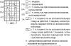
Types of abnormal location and placenta previa
In accordance with the specific features of the location of the placenta, specialists (note - based on information obtained after an ultrasound) distinguish certain types of placenta presentation.
- Full presentation. The most dangerous thing. An option when the placenta completely covers the internal os (note: the opening of the cervix). That is, the baby simply will not be able to get into the birth canal (the exit is blocked by the placenta). The only option for childbirth in this case is a caesarean section.
- Incomplete presentation. In this case, the placenta covers the internal os only partially (a small area remains free) or the lower part of the “child’s place” is located at the very edge of the internal os. In most cases, even with incomplete presentation, “classical” childbirth is also impossible - only a cesarean section (the child simply will not fit into part of the narrow lumen).
- Lower presentation. The most favorable option regarding the dangers of pregnancy and childbirth. In this case, the placenta is located 7 (or less) cm from the perimeter of the entrance directly into the cervix/canal. That is, the area of the internal os is not blocked by the placenta (the path “from the mother” is free).

The main types of placenta previa are low, complete and partial.
Symptoms and diagnosis of abnormal placental position - at what time can it be diagnosed?
One of the most “bright” symptoms of presentation is regular bleeding accompanied by painful sensations. It can be observed from the 12th week until childbirth - but, as a rule, it develops from the 2nd half of pregnancy due to severe stretching of the uterine walls.
In recent weeks, the intensity of bleeding may increase.
The following factors provoke bleeding:
Bleeding can be different, and the volume/intensity does not depend at all on the degree of presentation. In addition, it should be noted that bleeding can be not only a sign, but also a serious complication of presentation when it does not stop for a long time.
Symptoms of previa may also include:
- Circulating blood volume deficiency.
- Severe anemia.
- Hypotension.
- Preeclampsia.
And some indirect signs:
- High fundus of the uterus.
- Incorrect presentation of the fetus (note - gluteal, oblique or transverse).
In the 2-3rd trimester, the placenta may change its location due to its growth in the direction of the most blood-supplied areas of the myometrium. In medicine this phenomenon is called "placenta migration". The process is usually completed around 34-35 weeks.
Diagnosis of placenta previa - how is it determined?
- Obstetric external examination (note – height of the uterus, position of the fetus).
- Auscultation (with it, in the case of presentation, noise of the placenta/vessels is usually noted directly in the lower part of the uterus near the placenta).
- Gynecological examination with speculum. Palpation determines complete presentation if there is a soft and large formation that occupies all the vaginal vaults, and incomplete presentation - when it occupies only the lateral or anterior vault.
- Ultrasound. The safest method (compared to the previous one). With its help, not only the fact of placenta previa is determined, but also the size, area and structure, as well as the degree of abruption, hematoma and the threat of miscarriage.

The course of pregnancy with an incorrect location of the placenta and possible complications
Possible complications of the presentation of the “baby seat” include the following:
- Threat of miscarriage and gestosis.
- Maternal anemia and chronic fetal hypoxia.
- Fetoplacental insufficiency.
- Delay in fetal development.
It is worth noting that complete placenta previa in most cases ends in premature birth.
How does pregnancy proceed with established placenta previa?
- Period 20-. If the presentation is confirmed on the 2nd ultrasound, and there are no symptoms, then a regular examination of the expectant mother by her gynecologist-obstetrician is sufficient. Usually, additional drugs are prescribed to reduce the tone of the uterus. If there is even spotting, hospitalization is required.
- Period 28-32 weeks. The most dangerous period for both: with an increase in the tone of the uterus in its lower parts, the risk of detachment and serious bleeding increases with the small size and immaturity of the fetus. In case of marginal or complete presentation, a hospital is indicated.
- Period 34 weeks. Even in the absence of bleeding and severe fetal suffering, the expectant mother is advised to go to hospital until the birth. Only constant supervision by specialists can guarantee a successful outcome of pregnancy and childbirth.
Peculiarities of childbirth with incorrect location and placenta previa - is cesarean section always necessary?
With this diagnosis, childbirth can indeed be natural.
True, under certain conditions:
- Appropriate health status of mother and fetus.
- No bleeding (or its complete stop after opening the fetus/bladder).
- Regular and strong contractions.
- The cervix is completely ready for childbirth.
- Head presentation of the fetus.
- Minor presentation.
In what cases is a caesarean section performed?
- First of all, with full presentation.
- Secondly, in case of incomplete presentation in combination with one of the factors (several factors) : breech presentation of the fetus or multiple pregnancy, scars on the uterus, narrow pelvis of the mother, burdened obstetric history (abortions or miscarriages, operations, etc.), age over 30 years, subject to 1 birth.
- In case of persistent bleeding with a significant amount of blood loss (approx. – over 250 ml) and regardless of the type of presentation.
During a natural birth, the doctor first waits until labor begins (on its own, without stimulants), and after dilating the cervix by one or two cm, opens the fetus/bladder. If after this the bleeding does not stop or even increases in momentum, then an urgent cesarean section is performed.

Note:
Prevention of presentation, oddly enough, also exists. This - refusing or preventing abortions through the use of contraceptives and their correct use, timely treatment of inflammatory diseases and careful attention to women's health.
Take care of yourself and be healthy!
The website warns: self-medication can harm your health! The diagnosis should only be made by a doctor after an examination. Therefore, if you notice any alarming symptoms, be sure to consult a specialist!
Placenta previa refers to its unusual attachment in the lower region of the uterus. At the same time, it covers the internal os partially or completely, being located in the path of the emerging fetus.
Pathology occurs in 0.2-0.8% of cases of the total number of births. In multiparous women, the incidence of pathology increases to 75-80%.
If placenta previa is detected in the 2nd trimester, you should not worry too much, since with an increase in the growth of the uterus, the placenta can shift upward by 8-10 cm. This displacement is called “placenta migration.”
Reasons
There is no clear data on the occurrence of pathology. The most common reasons include:
- inflammatory diseases (for example, endometritis);
- a large number of births (more than 2-3);
- abortions;
- the presence of scars on the uterus, for example, after a cesarean section;
- uterine fibroids;
- abnormalities of the uterus.
Most of these reasons cause structural changes in the endometrium, which disrupt the normal attachment of the fertilized egg.
There are also fruit causes, which are determined by the characteristics of the fertilized egg itself.
Symptoms of placenta previa
The main and sometimes the only sign of placenta previa is uterine bleeding. It occurs suddenly, without other symptoms. More often it begins at 28-30 weeks, since at this time intense stretching of the lower part of the uterus occurs. Bleeding is characterized by its own characteristics:
- not accompanied by pain;
- scarlet color;
- often repeated;
- Anemia occurs in a pregnant woman.
Bleeding in the second trimester often indicates a complete presentation (completely covers the uterine pharynx), and at the end of pregnancy or the first stage of labor it is characteristic of an incomplete presentation (partially covers the pharynx). There is also a low presentation (the placenta is close to the uterine os).
During an obstetric examination, a gynecologist can reveal:
- painlessness on palpation of the uterus;
- breech presentation of the fetus;
- incorrect position of the fetus (oblique or transverse);
- absence of uterine hypertonicity;
- high standing of the presenting part of the fetus.
Using ultrasound, you can determine the degree of presentation:
- 1st degree: the edge of the placenta is located near the uterine os, at a distance of 3-5 cm (the most favorable option);
- 2nd degree: the edge of the placenta reaches the pharynx, but does not overlap it;
- 3rd degree: the edge of the placenta partially covers the uterine os;
- 4th degree: the placenta completely covers the pharynx.
With this pathology, to prevent bleeding, you need to avoid physical activity, air travel and maintain sexual rest.
If a pregnant woman experiences severe anemia or hemorrhagic shock, the child may experience acute hypoxia. Therefore, if blood is detected, you should immediately inform your gynecologist.
Placenta previa can be detected before bleeding occurs, since screening with ultrasound is mandatory in every trimester.
Diagnostics
Ultrasound is the most accurate and safe diagnostic method. All pregnant women undergo a screening test in each trimester, during which the location of the placenta is determined.
Treatment of placenta previa
Treatment is carried out in a hospital. The goal is to stop bleeding. If there is no bleeding and the placenta is low, then fetal hypoxia and developmental delay are prevented on an outpatient basis.
Non-drug therapy
- Bed rest is prescribed. It is recommended to exclude physical activity and sexual activity.
Drug therapy
- For minor bleeding, drug treatment is prescribed. Tocolytics (Hexoprenaline, Fenoterol), glucocorticoids (ascorbic acid 5% intravenously) and agents that strengthen the vascular wall (dexamethasone intramuscularly and orally) are used. The first and last group of drugs are used at any time. Glucocorticoids are prescribed up to 34 weeks to prevent neonatal respiratory distress syndrome (breathing difficulty caused by immature lungs in premature infants).
- Therapy is also carried out aimed at preventing anemia in a pregnant woman. During treatment, it is necessary to monitor the location of the placenta using ultrasound.
Surgical treatment
- If bleeding of 250 ml or more occurs, then an emergency cesarean section is indicated at any stage of pregnancy.
Delivery
In case of bloody discharge, delivery is carried out using a cesarean section.
In case of incomplete presentation and absence of bleeding at the beginning of labor, vaginal delivery is possible. But this issue is resolved individually and only by a doctor.
Possible complications with placenta previa
Complications that may occur with placenta previa include:
- Premature birth (20% of cases).
- Threat of miscarriage.
- Heavy bleeding.
- Anemia in a pregnant woman due to blood loss.
- Incorrect position of the fetus or breech presentation. It occurs because the placenta previa prevents the head from descending into the pelvis.
- Dense attachment of the placenta or true accreta (the placenta grows deep into the muscular layer of the uterus and thereby causes bleeding). This complication is rare, but usually ends with removal of the uterus.
- Chronic hypoxia and fetal development delay. It occurs due to reduced blood supply in the lower uterine segment.
- Detachment of the placenta previa. Accompanied by bleeding, emergency caesarean section is indicated. Most dangerous for the fetus.
Prognosis for mother and child
With minor bleeding and timely cesarean section, the prognosis is favorable. Placenta previa is a rather dangerous pathology, so it is recommended that you undergo all examinations (especially screening ultrasound) and strictly follow all the orders of your obstetrician-gynecologist.
If such a pathology is detected in the first or second trimester, you should not worry too much, as there is a high probability of “placental migration”.
Some studies during pregnancy
Low placentation during pregnancy is a serious complication that requires constant monitoring of the woman and emergency care if necessary.
The placenta is a temporary organ that forms by the second week of pregnancy, woven from the vessels of the mother and fetus. It attaches to the wall of the uterus, grows, develops and reaches maturity. Functions of the organ:
- saturation of the child’s blood with oxygen and removal of carbon dioxide;
- delivery of nutritional components to the fetus and removal of waste products;
- synthesis of hormones necessary for the normal development of pregnancy and preparing the female breast for milk production;
- immune protection of the baby in the womb.
The norm is that the placenta is attached to the posterior or lateral wall of the uterus. But if it is located too low, problems may begin.
Low placenta previa is an abnormality during pregnancy. It is characterized by attachment in the lower part of the uterus, which completely or slightly covers the internal pharynx. This means that complications are possible during pregnancy and childbirth.
Schematic representation of the problem
The condition often resolves spontaneously as the upper uterus moves out of place. This phenomenon is called placental migration. But in general, the risk of fetal death is quite high: from 7 to 25%.
The cause of the baby's death may be acute hypoxia due to insufficient placental blood supply or premature birth.
This pathology is also dangerous for a pregnant woman. Bleeding that occurs with placenta previa causes death in 1-3% of women.
The exact location of the attachment can be determined by ultrasound in the 3rd trimester. Normally, the location of the organ is located at a distance of 5 or more centimeters from the internal os of the uterus.
Reasons
Most causes of low placentation are due to diseases and conditions that occurred before pregnancy.
What causes deviation:
- inflammatory and infectious processes in the genital organs;
- damage to the uterine mucosa;
- previous miscarriages or abortions;
- gynecological interventions;
- multiple pregnancy. Women with twins or triplets are automatically at risk;
- birth by caesarean section;
- fibroids, endometritis and other diseases of the uterus;
- smoking, excessive alcohol consumption;
- many births;
- anomalies of the structure and development, functioning of the uterus;
- woman's age over 35 years.
The most common cause of low presentation is a previous uterine curettage. The procedure damages the mucous membrane, which prevents the fertilized egg from attaching to the upper part of the organ.

Consult a doctor
The insidiousness of deviation is that it practically does not manifest itself. Symptoms appear already at an advanced stage, when irreversible processes take place in the body, for example, exfoliation. These signs:
- heaviness in the lower abdomen, nagging pain;
- bloody discharge. When they appear, you need to call an ambulance;
- death of the fetus in the womb or its excessive activity due to hypoxia - lack of oxygen;
- severe toxicosis - 30% of women with this diagnosis suffer from it;
- In approximately half of the cases of pathology, ultrasound reveals a breech presentation of the fetus.
A pregnant woman herself cannot suspect an anomaly until clear symptoms appear. The condition is monitored during routine ultrasounds. The study allows not only to identify the problem, but to determine its extent and severity.
Types of low presentation depending on the location of the placenta:
- rear This arrangement of the organ is the most favorable option. In most cases, in later stages, the baby's place moves upward, freeing the birth canal. Pregnancy is relatively comfortable;
- front In this case, you need to prepare for difficulties. If the baby is large and active, he will put pressure on the placenta. This will cause problems with the umbilical cord, the risk of entanglement and compression. Such a presentation rarely changes before childbirth, which means that the birth canal will not be free;
- complete or partial, when the organ obscures the os of the uterus. Deviation requires great care. It is important to discuss all the details with your doctor before giving birth and prepare for the fact that you will have to give birth by cesarean section.
What to do
Diagnosis of low placentation allows one to determine how dangerous this condition is for the pregnant woman and the child. Conducted:
- analysis of symptoms: discharge, abdominal pain;
- Ultrasound is the main type of diagnosis, safe and informative. Carried out at 12, 19-20 and 30 weeks;
- bimanual examination of the vagina (provided there is no bleeding).
After confirming the diagnosis of low placenta previa, the doctor prescribes treatment and gives recommendations to correct the situation. If desired and competent actions are taken, the location of the organ can be changed.
If the pregnancy is proceeding normally, and the term has not reached 35 weeks, treatment is conservative. Strict bed rest, monitoring of the fetus and the intensity of bleeding are indicated. Any exertion or sexual contact is prohibited.

There are no medications that elevate the placenta. Medicines are prescribed to help improve the condition of the pregnant woman and promote migration of the baby's place. This:
- tocolytics, antispasmodics - stimulate stretching of the lower parts of the uterus;
- agents that reduce myometrial tone;
- iron-containing drugs - prescribed to women with bleeding to prevent iron deficiency anemia;
- medications that activate placental blood circulation - to avoid the development of hypoxia in the fetus;
- magnesia, intravenous glucose, vitamins.
The drug Utrozhestan helps prevent premature birth with low placentation. To prevent breathing problems in the fetus during childbirth, glucocorticosteroids are prescribed.
If the presentation is partial and accompanied by mild bleeding, conservative treatment helps to save the baby. But a woman must remember that at the slightest discharge and deterioration in health, she must immediately call an ambulance.
If there is heavy bleeding or the woman’s poor condition, the pregnancy is terminated for health reasons.
- severe blood loss (more than 200 ml);
- drop in blood pressure, anemia;
- complete presentation together with bleeding.

Ultrasound analysis image
The choice of delivery method if the pregnancy is carried to term depends on the indications. In case of complete presentation, the cervix of the uterus is closed, so a caesarean section is performed. It is also carried out when:
- placental abruption;
- polyhydramnios;
- incorrect positioning of the baby;
- scars on the uterus;
- multiple pregnancy;
- age after 30 years.
If the presentation is partial, natural childbirth is not excluded. But only if the baby is positioned head down, labor is active and the cervix is mature. In case of sudden bleeding, a puncture of the amniotic sac is performed. This helps stop the bleeding and lead to a normal birth.
If the cervix is not ready and the baby's head is small, a caesarean section is performed.
Why is it dangerous?

The consequences can be dire
Low placenta previa during pregnancy is a condition that is dangerous for both the woman and the fetus. Among the unpleasant consequences:
- the baby can damage the placenta with active movements. This is especially true for later stages, when a large baby puts pressure on the organ and is able to catch the placenta membrane;
- with low placentation, the cervix is not intensively supplied with blood, which is fraught with the development of fetal hypoxia;
- the anomaly threatens complications during childbirth, since the baby’s place prevents the baby from leaving the womb;
- insufficient placental blood circulation can cause increased activity of the fetus, which is fraught with twisting and clamping of the umbilical cord;
- Placental abruption is a dangerous condition that leads to the death of the fetus, and sometimes the woman. When complications occur, the stomach begins to ache and bleeding appears;
- Pregnant women with this diagnosis often experience low blood pressure and develop late gestosis.
The organ is densely intertwined with blood vessels connected to the uterus. This ensures placental blood exchange. And blood carries vitamins, proteins, oxygen, hormones and other substances necessary for life to the fetus.
With low presentation, the blood supply to the lower part of the uterus deteriorates. Consequently, the fetus is not fully fed with useful components. This increases the risks of intrauterine growth retardation and hypoxia.
If placental insufficiency and deterioration of blood flow are diagnosed, maintenance therapy is prescribed to compensate for the lack of nutrients in the fetus.
This is what low placentation threatens. A child can suffer from an anomaly at any stage of pregnancy. Caution and strict adherence to the doctor’s recommendations will help protect him and yourself from complications.
When it rises
Low placental presentation is detected at different stages. At the same time, it is difficult to predict the consequences, and it is unknown how long it will take to get up. Depending on the trimester of pregnancy, therapeutic actions to correct the pathology depend.
- 1st trimester. The first planned ultrasound is performed at 12-13 weeks, at which time the anomaly is detected. There is no need to worry during this period, since in 70% of cases the placenta rises by 20-21 weeks;
- 2nd trimester. By the period of twenty weeks, placental blood circulation is improved, but with low presentation it is disrupted, especially if the fetus is large and puts pressure on the organ from above. In this case, the gynecologist admits the pregnant woman to the hospital with strict adherence to bed rest and drug therapy. Usually by 22-23 weeks the baby's place rises up. If the situation does not change, the doctor gives the woman recommendations regarding lifestyle and continues to try to improve the situation;
- 3rd trimester. Mostly by 32-34.5 weeks the placenta moves upward under the pressure of the growing uterus. Then the problem disappears. If during pregnancy it does not come into place, at 36 weeks the issue of caesarean section is decided. This is especially important with complete presentation.

The cause could be curettage of the uterus
Depending on the trimester and the nature of the location of the placenta, different actions are taken to correct the anomaly. It is important for a woman to be patient and not panic.
In most cases, the placenta rises towards the end of pregnancy. If this does not happen, but the woman and baby feel well, a caesarean section is performed.
What not to do
Pregnant women with low placentation must be constantly monitored by a doctor. If you follow his recommendations exactly, everything will be fine. What you shouldn't do:
- worry. Modern medicine successfully treats pregnant women with low placental attachment. In 90% of cases, a woman gives birth to a healthy baby. Moreover, 60% of births are carried out naturally, and only 40 - by cesarean section;
- have sexual intercourse. Sex at any stage can damage the organ and lead to detachment. This only applies to women with low placenta previa;
- play sports, do abdominal exercises, lift weights, walk a lot. Decide what is more important, an active lifestyle or the health of the child;
- do douching and any other vaginal manipulations so as not to harm the pregnancy;
- worry, get nervous, get irritated. This will lead to an unhealthy emotional atmosphere and aggravate the condition. Develop stress resistance;
- travel on public transport, visit places with large crowds of people. They can push there, which will cause even greater prolapse of the organ;
- Ignore the doctor's recommendations and do not go to the hospital when necessary.

You have to be patient
If the presentation is low, the woman is advised to place a pillow under her legs so that they are higher than the level of the body. This will help the placenta quickly find its place.
Low placentation is not a disease, but a special condition. The situation requires, first of all, not treatment, but correction. Much depends on the mood of the pregnant woman, her actions, and how accurately they correspond to the advice of doctors.
Preventive measures will help to avoid anomalies. These include:
- prevention of infectious and inflammatory diseases, their timely treatment;
- maintaining a healthy lifestyle: proper nutrition, adherence to work and rest schedules, not abusing alcohol and tobacco;
- protection against unwanted pregnancy, so that there is no history of abortion;
- performing a caesarean section only in cases where there are vital indications;
- performance of gynecological manipulations and operations in trusted clinics by experienced doctors.
Thank you 0
What is the danger and how does childbirth take place?
During pregnancy, the placenta becomes the organ that connects two organisms (the expectant mother and her developing baby) into a single whole. Special cells of the outer embryonic membrane of the fetus take part in its formation, which in very early stages of pregnancy produce enzymes that help the embryo penetrate the uterine wall.
In the vast majority of cases, the placenta is attached in the upper part of the uterus, in the area of its fundus - in this position and in the absence of other pregnancy complications, optimal conditions are created for the development of the fetus.
Important differences
It is worth clearly distinguishing between low location of the placenta during pregnancy (low placentation) and placenta previa.
In the case when an ultrasound examination reveals the location of the placenta in lower parts of the uterus, but at a distance of at least 5 cm from her internal os, the doctor diagnoses low placentation during pregnancy. If the placenta is partially or completely covers the area of the internal pharynx, then this condition is called, respectively, complete or partial placenta previa.
Placenta previa occurs in less than 1% of all pregnancies. Of these, about 1/3 is complete placenta previa and 2/3 is partial.
With a low-lying placenta, unlike previa, the placental tissue never comes into contact with the cervix and, even under the most unfavorable circumstances, does not interfere with the normal course of natural childbirth.
Causes
Gynecologists note that the shorter the gestational age, the greater the likelihood of diagnosing low placentation. It has been proven that the placenta has a unique ability to migrate.
The essence of the phenomenon of placenta migration is that with further progression of pregnancy, the placenta can independently move to areas of the uterus with a well-developed network of blood vessels.
That is why, even if during the first ultrasound examination of a pregnant woman, usually carried out at 11-12 weeks of gestation, the doctor discovers that the placenta is forming in the lower part of the uterus, one should not despair - the placenta moves weekly and by the time of delivery this situation may disappear on its own.
The causes of placenta previa are:
- various inflammatory processes affecting the mucous membrane of the uterine body - much more often this condition is observed during repeated pregnancies (both those that ended in termination at an early stage, and those complicated by purulent inflammation of the uterus in the postpartum period);
- surgical manipulations affecting the uterine mucosa - diagnostic curettage (even performed when indicated), abortions, therapeutic hysteroscopies, during which polypectomies were performed;
- neoplasms of the uterus - polyps, fibroids, fibroids, especially when they reach significant sizes;
- multiple pregnancy;
- malformations of the uterus – saddle-shaped deformation, bicornuate uterus.
Actually, the main prevention of low placentation, carried out before pregnancy, is the prevention of abortion, performing curettage only for strict health reasons, as well as timely detection of malformations.
How does a low placenta manifest and what does it mean?

Until the moment when the placenta, as it develops, reaches the internal uterine os, there may be no clinical manifestations of this condition - the pregnant woman has no complaints at all, and low placentation is diagnosed only during an ultrasound examination.
It is for the timely detection of pregnancy complications, including the detection of features of the location of the placenta, that every pregnant woman should, while waiting for the child, attend three mandatory ultrasounds(at 11-12 weeks, at 20-21 or 22-24 weeks, at 32-33 weeks of normal pregnancy).
Each pregnancy is individual, so there are no norms for the location of the placenta by week. It is important to undergo ultrasound in a timely manner and carefully follow the advice of your gynecologist.
Even if during the first ultrasound the doctor detects a tendency towards low placentation, in the absence of the woman’s complaints and symptoms of a threatening miscarriage (increased uterine tone, bleeding from the genital tract, pain in the abdomen and lower back), no treatment is prescribed. If a woman develops any of these signs, the same treatment is prescribed as for a threatened miscarriage.
During pregnancy, it is important to pay attention to the appearance of signs of any, even minimally expressed bleeding - if it occurs, the expectant mother must be hospitalized in the gynecological department or the pregnancy pathology department.
If a low level of placenta is detected, it is necessary to attach the placenta limiting physical activity for a pregnant woman– any work related to heavy lifting is prohibited (if necessary, an appropriate conclusion from the Higher Quality Commission is issued for transfer to light work), a reduction in physical activity is recommended and sexual activity is limited until the threat of miscarriage disappears.
How does childbirth go?
It has been proven that in the vast majority of cases, by the 35-36th week of pregnancy, low placentation spontaneously stops and by the time of delivery the placenta takes its normal position on the anterior or posterior surface of the uterus. Actually, a doctor can make a diagnosis of “placenta previa,” which is an absolute indication for a planned cesarean section, only by this date.
If low placentation disappears and the woman does not have any other indications for surgical delivery via cesarean section, then there is no need to refuse vaginal delivery.
You might also be interested in:

Gemma is an example of miniature carving of colored stones and gems - glyptics. This view...
98/104 (110/116) 122/128 You will need Yarn (100% cotton; 125 m / 50 g) - 250 (250) 300...

Periodically replenishes its collection of publications dedicated to various colors and shades in...

A correctly tied scarf around the neck affects the external image and characterizes the internal...

251 03/26/2019 6 min. You can wear high heels and an expensive suit, make...

