


There is not a single woman in the world who would not dream of becoming a mother. But not every pregnancy can bring only positive emotions and feelings. What to do if you heard such a terrible word from a gynecologist that leaves you in shock? What are the main signs of an ectopic pregnancy in the early stages? What to do so that it is not too late?
Characteristics of an ectopic pregnancy
An ectopic pregnancy is a condition that is dangerous to the life and health of a woman. Such a pathology can occur if, after fertilization of the egg, the embryo was unable to attach to the uterine cavity, but stopped in the fallopian tube and began its active development there. The reasons for this problem are written below, but it also happens that they simply do not exist. It is very important to know that if such a disorder has already been present, then the next pregnancy has a chance of being the same.
The main types of ectopic pregnancy in the early stages
Once the early signs of an ectopic pregnancy are identified, its type can be easily determined. Today, there are three types of such pathology: ovarian, tubal and abdominal. If an ectopic pregnancy is observed (5 weeks), then it entails an enlargement of the uterus, as well as the beginning of the preparation of the mucous membrane to fully accept the embryo. After such a period, it will be quite difficult to establish the real fertilization period, since the size of the uterus will not correspond to reality.
In the early stages, tubal pregnancy most often develops. It is disrupted externally and internally, that is, by complete rupture of the fallopian tube. If this happens, the woman will begin to bleed very heavily. This process will also be accompanied by severe pain, which can lead to loss of consciousness. This condition is often accompanied by pale skin, increased sweating, dilated pupils, dry mouth, decreased blood pressure, and a weak pulse. Removal of an ectopic pregnancy is carried out as a tubal miscarriage. In this case, the patient experiences a slightly elevated body temperature, abdominal pain and dizziness. Fainting conditions were also observed. The pressure continues to be normal, and after a couple of days, bloody discharge will be visible from the vagina.
When an abdominal pregnancy occurs, it is very difficult to recognize an ectopic pregnancy. But over time, adhesions will begin to appear in the peritoneum, which will cause severe pain, constipation and vomiting. After such a pathology, doctors will diagnose “late miscarriage,” which begins at the initial stage. The cervix will remain completely closed.
The development of ovarian pregnancy begins in the embryonic horn of the uterus. It is most often observed in women who have pathology of the development of the uterus.
After the first signs of an ectopic pregnancy are noticed, the patient must be taken to the hospital and have surgery.
Tests that detect ectopic pregnancy

How to determine an ectopic pregnancy so that in the future it does not end in tragedy? There are several ways. When analyzing hCG blood, this pathology can be recognized. If the indicator is significantly lower than expected at the current stage, then these are the first signs of an ectopic pregnancy, which you need to pay special attention to.
This problem can also be identified by ultrasound, since it can be seen where the fetus is located, in the uterus or not. If there is a suspicion of an ectopic pregnancy, then it is best to examine it with a transvaginal ultrasound, since the sensitivity of this method is much higher, and the probability of recognizing the pathology is high, even at short notice.
Does the fetus survive an ectopic pregnancy?
This type of pregnancy can never result in the successful development of the fetus; in any case, it dies. The sooner treatment is started and the woman receives first aid, the higher the chances of avoiding complications and preserving the patient’s fallopian tube.
Why is an ectopic pregnancy dangerous?
This pathology is considered very dangerous, as it can cause enormous blood loss, infertility, and in the future even death. If you have the first signs of an ectopic pregnancy in the early stages, then you need to be under the strict supervision of doctors who will be able to provide qualified assistance in a moment of crisis.
Signals for concern

There is a list of main symptoms at which you need to start worrying about your health. These include: general malaise, sometimes fainting and constant dizziness. It should also be taken into account that the older a woman is, the more likely an ectopic pregnancy is. Symptoms, the timing of which is short, in girls after 35 years of age should be subject to increased attention from specialists. For women at risk, it is recommended to undergo an ultrasound every two weeks so that the possibility of pathology can be excluded. This is done until the fertilized egg is clearly visible.
Diseases after which ectopic pregnancy develops
Transferred diseases very rarely pass without a trace on the body, so there are also those that are fraught with the appearance of an ectopic pregnancy. These include:
Operations performed on the organs of the reproductive system;
Laparoscopy;
The use of drugs "Postinor", "Escapelle", as well as other similar drugs.
Signs of an ectopic pregnancy in the early stages may not always be clearly expressed, since diagnosis still remains very difficult, and a pregnancy test cannot indicate the problem at all.
Symptoms of an ectopic pregnancy

At the initial stage, it is quite difficult to recognize the occurrence of an ectopic pregnancy. It all starts like everyone else, there is a delay in menstruation, increased tenderness of the mammary glands, toxicosis, changes in appetite and drowsiness.
After 5-8 weeks have passed, everything changes dramatically, and the first signs of an ectopic pregnancy begin to appear. A woman increasingly notices huge, severe cutting and aching pain in the lower abdomen, usually on the side on which the egg is attached. If a pregnant woman has a hemorrhage in the peritoneum, she will most likely experience pain in the anus, as well as during bowel movements and urination. The pain syndrome is accompanied by bloody discharge, which is completely unrelated to the blood in the peritoneum.
At this point, the body begins to exhibit such a reaction due to a decrease in the level of progesterone, which is the main hormone of pregnancy. In such a situation, the woman is in a state of shock.
The passage of an ectopic pregnancy at different stages

Ectopic pregnancy in the early stages initially manifests itself as severe vaginal bleeding, and women often complain of significant pain in the lower abdomen. They are much stronger than during a normal pregnancy. Severe and very frequent attacks of vomiting and nausea appear, as well as discomfort when urinating and defecating. If these symptoms appear, you should urgently contact your doctor, with whom the woman is registered.
Late periods are accompanied by pain in the neck and shoulders. This is due to the fact that at this stage the fertilized egg becomes larger and hemorrhage begins in the abdominal cavity. On an emotional level, there is increased fatigue, a huge feeling of apathy and irritation, which is much brighter than during normal pregnancy.
If a woman during her ectopic pregnancy complains of severe sweating, then this should indicate a huge risk to her life.
High risk of ectopic pregnancy
The risk increases in the following cases:
For girls who have many non-regular sexual partners;
In women over 35 years of age;
If pregnancy occurs with an intrauterine device;
For those who have suffered from sexually transmitted diseases in the past, even if they have completed a full course of recovery and there seem to be no symptoms, the risk still remains high;
If conception occurred after a tubal ligation procedure;
In patients who have undergone operations on the intestines and pelvic organs in their lives;
All those who took the drugs "Postinor" or "Escapelle"; they remain at risk for ectopic pregnancy for a month after taking the drugs;
In the event that a woman could not get pregnant for several years (at least 2), the risk of such a pregnancy is very high;
For those who smoke regularly.
What should a woman do if she has an ectopic pregnancy?
If, after all the first tests, ultrasounds and studies, your doctor diagnosed an “ectopic pregnancy”, what should you do when your thoughts are confused and you don’t understand anything? Of course, from this moment on, the patient must be constantly under the close supervision of doctors. Treatment can take place in three main areas:
Waiting tactics - after an ectopic pregnancy has been determined, the duration of which is still short, the woman is constantly under the supervision of doctors, but nothing is done;
Drug treatment is therapy with drugs, but without surgery;
The operation involves removing the fertilized egg, and in severe cases, the fallopian tube as well.
How to recognize a ruptured fallopian tube

This phenomenon can occur at very advanced stages, when a woman does not know how to identify an ectopic pregnancy. Subsequently, the embryo becomes very large, and under such influence the fallopian tube cannot withstand it and bursts. This condition is considered very dangerous for a woman, since subsequently everything can end in death. The latest period at which an ectopic pregnancy can terminate on its own is considered to be 9-10 weeks. If, during the pathology, blood is discharged without stopping, the pressure decreases, and there is clouding in the eyes and a state of fainting is felt, that is, all the signs of an ectopic pregnancy appear in the early stages, then it is necessary to urgently call an ambulance for emergency hospitalization. This condition may be accompanied by acute abdominal pain.
Life after pregnancy and its consequences

All the horrors are left behind, the operation is completed, and the patient begins to return to normal life. Then the woman undergoes a course of treatment with antibiotics, she continues to be injected with painkillers, as well as vitamins that support the body.
With the help of physiotherapeutic procedures and drug treatment, the patient endures this period easier, the condition of the fallopian tubes begins to improve, and recovery is accelerated.
In order to avoid problems with pregnancy later in life, the woman is prescribed a course of anti-inflammatory drugs. In the event that a tubectomy was performed, namely the removal of the fallopian tube, then its remainder should also be treated. After such a pathology, the patient may encounter late complications. After a tubectomy, there is a possibility of infertility. There also remains a very high possibility of recurrence of ectopic pregnancy, namely 15%.
Contraception should be one of the main and primary places, since after such a pregnancy you cannot plan the next conception for 3 months, but it is best to wait six months. It is very important that planning is discussed with the doctor who monitors the woman’s tests. It will be clear from them whether the body is ready for new loads or not. Initially, you need to examine the patency of the fallopian tubes. It is also worth paying close attention to the diagnosis of inflammatory processes and infections. If all tests show positive results, then the woman, on the recommendation of her doctor, can think about planning a new pregnancy.
The diagnosis of ectopic pregnancy (hereinafter referred to as EP) sounds very unpleasant. In modern medical practice, such cases are recorded quite often. The condition is not only accompanied by pain and many other unpleasant symptoms, but also poses a serious threat to the patient’s life. To prevent dangerous consequences, every woman should know the signs of an ectopic pregnancy in the early stages. You can read about them in detail in the article.
What does the term ectopic pregnancy mean? When an egg is fertilized in the fallopian tube, it moves to the uterus, where it is fixed and has all the necessary conditions for the normal development of the fetus. In the case of an ectopic pregnancy, the egg does not enter the uterus, but is implanted elsewhere. The most common type of ectopic pregnancy is localization in the fallopian tube. Less commonly, it happens that a fertilized egg begins to develop in the peritoneal cavity, on the cervix or the ovary itself.
Early signs
How to determine a pathological pregnancy? In the early stages, it is quite difficult to recognize an ectopic pregnancy. The fact is that every girl’s body is individual and the signs of such a pregnancy can manifest themselves in different ways. So, what manifestations may indicate the fixation of a fertilized egg outside the uterus? Let's figure it out.
Increase in basal temperature
Many representatives of the fair half are accustomed to monitoring their basal temperature (BT). By measuring the temperature in the anus, you can determine the onset of ovulation or early pregnancy. Due to the increased amount of progesterone, BBT will remain high throughout the cycle. If conception does not occur, BT decreases. It is worth noting that this indicator can be used to determine the onset of pregnancy in general, but not specifically ectopic conception.
Lack of menstruation
Lack of menstruation is the surest way to determine early conception. Despite this, we must not forget that other reasons may also lead to a delay. These may be provoking factors such as nervous shock, hormonal imbalance, serious illnesses, including oncology. In addition, you cannot be one hundred percent sure that there is no pregnancy when menstruation occurs, since many girls experience menstruation even after conception.
Morning sickness
Early pregnancy can be recognized by morning sickness. Many women know about this ailment when they become pregnant. Often this symptom also appears in the daytime and evening. This is due to hormonal changes in the body, a drop in sugar and some other reasons. If you notice such signs, you should take a pregnancy test. The most accurate test is considered digital. If it shows two stripes, it is important to undergo a medical examination as soon as possible.

If an ectopic pregnancy occurs, it will be possible to diagnose it early and prevent complications.
Changes in the mammary glands
When pregnancy occurs, many girls already in the early stages notice the following signs from the breast:
- darkening of the nipples, increasing their sensitivity;
- painful sensations in the chest, it becomes as if lumpy;
- breast swelling;
- veins in this area become clearly visible;
- darkening of the areolas;
- bumps on the areolas become more noticeable, and their number often increases.
It happens that some of these manifestations, for example, soreness, occur at the beginning of menstruation. This should be taken into account when assessing such changes.
Increased vaginal discharge
Normally, girls produce cervical mucus. When pregnancy occurs, the concentration of the hormone progesterone increases in the body. In this regard, a woman may already feel in the early stages that the amount of mucus has increased. Not all girls may notice this sign. For many women, the amount of secretion may be completely insignificant.
Fast fatiguability
When pregnancy occurs, no matter whether it is ectopic or normal, the woman’s body undergoes various changes already in the early stages. At the same time, metabolic processes are enhanced to maintain normal fetal development. This often leads to rapid fatigue. Girls note a constant desire to sleep, and physical activity is significantly reduced. In addition, the hormone progesterone, which has a sedative effect, acts as a relaxing factor. All this leads to loss of ability to work, a constant desire to lie down and sleep.
Frequent urination
Another common sign of early pregnancy is increased urination. Already 7-10 days after conception, a girl may experience such a phenomenon as frequent urination. The fact is that when pregnancy occurs, the body begins to produce human chorionic gonadotropin. HCG is the so-called pregnancy hormone. As a result, the blood supply to the pelvic organs increases. In this case, the urge to urinate is observed even with a slight filling of the bladder. This symptom intensifies at night.
Painful sensations in the lower abdomen
Will my stomach hurt during a pathological pregnancy? Spasms are often observed with the onset of ectopic fetal location in the early stages. This is due to the fact that the fetus grows and develops. Cramps can also occur during normal pregnancy. In this case, painful sensations are considered quite normal, because the uterus is constantly contracting, and spasms are observed during the development of the fetus.
The alarm should be sounded if the pain is accompanied by bleeding. A sign such as the appearance of blood may indicate both an ectopic pregnancy and a threat of miscarriage. In such a situation, you need to contact a gynecologist as soon as possible. If we consider the question of when pain begins during an ectopic pregnancy, it should be noted that most often a woman experiences discomfort when the embryo reaches about 5 cm in diameter, that is, at 12-16 weeks.

Many girls experience cramps during menstruation. These two conditions should not be confused. Every woman knows when she menstruates and what the nature of the discharge is.
Indigestion
Due to hormonal changes, the normal functioning of the intestinal tract is disrupted. The walls of the stomach and intestines relax under the influence of progesterone, and peristalsis decreases. This can lead to excessive gas, constipation or, conversely, diarrhea and other symptoms. Such manifestations often indicate the onset of pregnancy, including ectopic pregnancy.
Increased sensitivity to odors
Another early sign of conception is a change in the acuity of smell. Girls are keenly aware of all smells. Many women find it difficult to cope with even everyday cooking. Any smell can cause disgust.
Nasal congestion
When conception occurs, immunity decreases. This is a normal physiological process that ensures the body accepts the fetus. At the same time, many girls face colds and nasal congestion. In addition, swelling of the nasal passages is also associated with changes in hormonal levels.
The appearance of acne
Another early sign of pregnancy is the formation of acne and pimples on the body. Of course, this is not a mandatory sign of conception, but many girls who have not previously suffered from acne may experience this phenomenon. Along with inflammation, acne may appear on the skin.
Change in taste preferences
Due to hormonal changes, some ladies may experience different tastes in their mouth. Often girls talk about the appearance of a metallic taste. In addition, stories about the desire to eat completely incompatible foods during pregnancy are far from myths.
Emotional outbursts
Emotional disturbances are a common early sign of pregnancy, including ectopic pregnancy. In this case, the mood can change several times a day. A woman can laugh and then cry five minutes later. Such “swings” are observed due to changes in the hormonal balance of the body.
Positive pregnancy test
And, of course, the surest way to determine early pregnancy, which can be done at home, is a test. Will the test show whether an ectopic pregnancy has occurred or not? Today there are a lot of such tests. Many of them accurately determine the increase in hCG from the first weeks of conception. If you see 2 clear stripes or one fuzzy line and the other a clear line, you should rush to see a doctor. From the first days of conception, the test may show a negative result.

The earlier an ectopic pregnancy is diagnosed, the greater the chances of maintaining health and eliminating dangerous consequences.
Signs of a tube rupture during an ectopic pregnancy
Organ rupture occurs when the patient does not go to the hospital in a timely manner. If a girl has not undergone the necessary examination, including ultrasound, screening and other methods, an ectopic pregnancy may remain undiagnosed and lead to very negative consequences. One such complication is a ruptured tube followed by internal bleeding. The fact is that only the uterus has a special structure. This organ is capable of stretching, which is a necessary condition for carrying a pregnancy. In contrast, the fallopian tubes are made of muscle fibers and dense tissue. They cannot stretch, so rupture occurs as the fetus develops.
Signs of a rupture:
- acute pain on the right or left side of the lower abdomen;
- cold, sticky sweat;
- a sharp drop in blood pressure;
- nausea, repeated vomiting, not leading to relief;
- headache and dizziness;
- increased heart rate;
- weakness, fainting.
If you try to answer the question of how long it takes for an organ to burst, you will hardly be able to give an exact answer. More often this happens in the period from the second to the third month. But cases of rupture are often recorded during the period from the fourth to the sixth week of ectopic pregnancy. This is due to several factors. For example, it depends on the size of the organ, the speed of fetal development, the presence of deviations in the formation of the embryo, and so on.
If such signs are detected, you should call an ambulance as soon as possible. In a clinical setting, with timely treatment, it is usually possible to save the patient’s health and life.
Why does the egg implant outside the uterus?
Why does the syndrome occur - ectopic pregnancy? As we have already found out, unlike intrauterine, with ectopic the fetus is attached in most cases in the fallopian tube and only in 5% of cases in the abdominal cavity or ovary. In this case, fertilization of the egg by the sperm occurs in an absolutely natural way.

The reasons for this violation are very diverse. Let's look at the most common of them.
Physical disorders in the body
Due to various diseases affecting the female organs, a fertilized egg may linger outside the uterus. The most common causes of ectopic pregnancy are the following conditions in women:
- adhesions;
- cysts;
- scar formation;
- oncological diseases of the reproductive system;
- undergoing an abortion;
- various inflammatory processes;
- sexual infections.
To prevent the formation of adhesions and scars, you should visit a female gynecologist every six months. In the early stages, such diseases are successfully treated with medication and minimally invasive surgical techniques.
Congenital diseases of the uterus and appendages
The etiology of ectopic pregnancy often lies in the presence of congenital diseases of the female reproductive system. Often the patient learns about congenital pathologies after an ectopic pregnancy has been diagnosed. If it is possible to identify this dangerous condition in a timely manner, early treatment, as a rule, is carried out without negative consequences for the patient’s life.
Some types of contraception
No matter how it sounds, some contraceptives can actually cause ectopic pregnancy. The fact is that they protect the uterus from the attachment of a fertilized egg, but do not act in the same way on other parts of the reproductive system.
If a woman uses a spiral, it is important to remember that this type of protection lasts no more than five years. If it stays in the body longer, the spiral no longer fully performs its functions. In this case, the fetus can attach to the cervix.
Artificial insemination
Artificial insemination is a method that allows a woman to become pregnant who cannot do so naturally for various reasons. It would seem that this procedure should completely eliminate any risks. Despite this, according to medical statistics, cases of ectopic pregnancy during IVF are observed quite often. The doctor is obliged to warn the couple who decide to conceive a child in this way about such risks.
Other reasons
In addition to congenital and acquired diseases of the female organs, taking contraceptives and IVF, one can identify factors that provoke cases of ectopic attachment of a fertilized egg. Pathogenesis includes:
- hormonal disorders of the body;
- smoking. This is due to the fact that nicotine reduces the level of female hormones;
- treatment with douching;
- woman's age after 30 years.
Bad habits are a common cause of ectopic pregnancy.

Experts draw attention to the fact that patients who have experienced an ectopic pregnancy once are much more susceptible to the likelihood of a recurrence of this pathological condition.
Types or classification of WB
In medical practice, ectopic pregnancy is classified according to the location, that is, the attachment of the fertilized egg, and according to the stages of its course. The following types are distinguished by location:
- in the fallopian tubes. The ICD-10 code is O00.1. This type is considered the most common, occurring in more than 90% of all women. In this case, the embryo is attached to the tube, where it grows and develops until it bursts or a spontaneous miscarriage occurs;
- in the ovary (ovarian attachment). The ICD-10 code is O00.2. The attachment of an egg in the ovary is diagnosed extremely rarely (1-2% of cases). This condition is observed due to the entry of a fertilized female cell into the ovary. The danger is that the organ bursts due to the growing embryo;
- in the abdominal cavity (sebarami abdominal pregnancy or Manga pregnancy). The ICD-10 code is O00.0. Here the fertilized cell attaches to one of the organs of this cavity. Medicine has recorded cases where, with such an attachment, pregnancy proceeded normally, but in most cases severe defects in the fetus are still observed;
- in the area between the large ligaments of the reproductive organ. This is a disrupted intrauterine pregnancy. If it is not interrupted in the early stages, the fetus can develop quite normally up to twenty weeks;
- in the area of the cervix (cervical attachment). In the early stages, there are no clinical signs of conception and attachment of the fetus in the cervix. Later, a woman may notice bloody discharge and pain during sexual intercourse;
- in the rudimentary uterine horn. This type of pathological condition is accompanied by spontaneous abortion no later than 4 months after cell attachment and heavy bleeding. The danger lies in significant blood loss; a woman can die within a few hours;
- interstitial appearance or attachment in the interstitium of the fallopian tubes. As with other types of pathological attachment of the egg, there are no signs in the early stages of the interstitial type. Pregnancy can proceed absolutely normally up to twenty weeks.
With any type of VD, the chances of carrying and giving birth to a healthy child are reduced to almost zero. The maximum duration of an ectopic pregnancy is considered to be up to 20 weeks, with the exception of the attachment of the fetus in the abdominal cavity. Only with an abdominal location is there a slight chance of normal fetal development.
Such branches of medicine as obstetrics and gynecology deal with ectopic pregnancy. To make diagnosis easier, doctors divide VD into several stages of development.

The dynamics of development may be as follows:
- Progressive pregnancy (early stage).
- Pregnancy under threat of termination (interrupted). This is a condition in which the fallopian tube bursts or spontaneous rejection of the embryo occurs;
- Completely interrupted ectopic pregnancy.
In addition, there is a condition in which there is a pathological attachment of two fertilized eggs in different parts at once or a multiple pregnancy, during which one of the fertilized eggs is attached to the uterus, and at the same time another cell is attached outside of it.
Is it possible to conceive a child after an ectopic pregnancy?
On numerous forums you can find questions from women about whether it is possible to get pregnant normally and give birth to a healthy child after VB? Let's try to answer this question.
When should you plan for another pregnancy?
How long before you can get pregnant after VB? Sufficient time must pass after the operation to remove the pathologically located egg. This is necessary for postoperative sutures to heal normally. In addition, it is necessary to carry out therapy aimed at preventing the formation of tissue scarring. After 6 months, the specialist should prescribe the patient a thorough medical examination, including ultrasound examination, histology analysis, etc. If the wounds have healed normally and there are no other contraindications, the woman is allowed to conceive a child.
If during the period of VB a rupture of the right or left tube occurs, the chances of successfully conceiving a child are halved, but a woman can still become pregnant and give birth.
Possible difficulties
Often, when the fallopian tube ruptures, doctors decide to completely remove it. This is due to the fact that the risk of re-pathological attachment of the fertilized egg in this area is quite high. Despite the surgery and the absence of one of the tubes, the patient may well become a mother. To do this, you need to undergo competent rehabilitation, listen and follow all the recommendations of doctors. Even if the expected conception does not occur immediately, do not despair. The methods of modern medicine can solve many problems with conceiving and bearing a child.
Finding out that ectopic implantation of the fertilized egg has occurred in the early stages is quite difficult. This can only be checked in a hospital. As a rule, this condition is detected when the first signs appear, which coincide with the symptoms during normal pregnancy. The patient notes the absence of menstruation, toxicosis, change in taste, worsening of smell, drowsiness; ultrasound can detect changes in the endometrium. If you notice such signs, you should immediately visit a female gynecologist. Unfortunately, not a single patient is insured against ectopic implantation of a fertilized egg. If you suspect this dangerous condition, you should not hesitate; you must make an appointment with a doctor as soon as possible.
Differential diagnosis
To distinguish VD from other pathologies, a protocol is drawn up, its differential diagnosis with appendicitis, ovarian apoplexy and other diseases is carried out. If doctors detect a ruptured tube or a frozen pregnancy, the woman is indicated for urgent surgical intervention. The most accurate and informative method used in making an early diagnosis of VD is ultrasound.

Using an ultrasound examination, you can detect a right-sided or left-sided pregnancy, as well as see the period of development.
Puncture
Using a puncture, the doctor can confirm or refute the diagnosis of ectopic pregnancy. This method is called Douglas space puncture. This space is the section of the abdominal zone between the anterior surface of the rectum and the uterus. By piercing the posterior fornix of the vagina, the doctor obtains fluid, which is examined in laboratory tests. Decoding the results allows you to get an accurate diagnosis.
Does the hCG level increase during VB?
The implantation of an egg outside the uterus also entails the production of hCG. This is an absolutely normal process that is observed in every woman during pregnancy. This test can be done at any clinic. Sometimes women wonder if this indicator can fall during VB? In fact, the level of the hormone during ectopic pregnancy may be slightly lower than during normal pregnancy, but it begins to fall only after the twelfth week. How the hCG level changes during VB can be seen in the table.
| Week of pregnancy | Human chorionic gonadotropin level |
| From 1 to 2 | 5,7-750 |
| From 3 to 4 | 150-7 200 |
| From 4 to 5 | 1000-31 900 |
| From 6 to 7 | 3 500-160 000 |
| From 7 to 8 | 64 000-155 000 |
| 8 to 9 | 460 000-211 000 |
| From 10 to 11 | 130 500-63 000 |
| From 11 to 12 | 13 500-63 000 |
| From 13 to 14 | 12 000-71 000 |
| From 15 to 25 | 8 000-60 000 |
| From 26 to 37 | 5 000-55 000 |
The table shows that at week 12 the hCG level reaches its maximum value, after which it begins to fall.
Corpus luteum in VB
After ovulation, a temporary gland is formed in the ovary, the role of which is to produce progesterone. If fertilization does not occur 2 weeks after the release of the egg, the corpus luteum dies. If conception occurs, this gland does not die, but continues to produce progesterone. If 2 weeks after ovulation there is no menstruation, and pregnancy is not visible on an ultrasound, conception can be determined by the presence of this corpus luteum. That is, the presence of this gland 14 days after ovulation means the onset of pregnancy, including ectopic pregnancy.
Use of laparoscopy
Modern techniques such as laparoscopy make it possible to identify and eliminate the pathological condition in the early stages. This minimally invasive technique is used not only for diagnosis, but also for embryo removal. Laparoscopic technique is an effective, but at the same time safe method for diagnosing and treating VB.
What does an ectopic pregnancy look like in a photo?
Some patients are interested in whether VB is visible in the early stages on ultrasound? Yes, it is indeed possible to recognize the condition using ultrasound. In the pictures you can see places of pathological attachment of the fertilized egg in various areas.

As already mentioned, pipe fastening is the most commonly diagnosed problem.
Treatment methods
What to do if you suspect you have an ectopic pregnancy? The question of how to remove this condition is relevant for many women. First of all, you should contact a gynecologist. The doctor will order the necessary tests and select the optimal treatment method. Without proper medical care, there is a serious threat to a woman’s life and health.
Drug treatment
Treatment of VB with conservative therapy is carried out only in the early stages in quite rare cases. In this case, micropreparations and macropreparations are used. This type of therapy often involves the use of the drug Methotrexate. This remedy leads to the freezing of the embryo and its resorption. The main disadvantage of this treatment is the risk of systemic side effects. Therefore, it is recommended to treat VB with this medication only when necessary. In addition, the drug Duphaston is used. This is a hormonal drug that increases the level of progesterone in the blood.
Laparoscopy
Laparoscopy is a low-traumatic surgical technique used for the treatment and diagnosis of many diseases of human internal organs. The operation is performed under general anesthesia, so there is no discomfort during the procedure. In addition, laparoscopy is performed with minimal damage to the patient’s tissues, as a result of which rehabilitation takes a short period of time.
How is the operation performed? The technique is carried out in several stages:
- The operated area is treated with an antiseptic.
- A small incision is made on the left or top of the umbilical area.
- A Veress needle is inserted, the purpose of which is to introduce carbon dioxide into the abdominal cavity.
- A laparoscope (a device with a camera) is inserted.
- Through additional incisions, instruments are inserted, with which the surgeon removes the embryo.
During the operation, the doctor decides whether to remove or preserve the fallopian tube. If it is subject to irreversible changes, it is removed along with the attached egg. The advantage of the technique is good efficiency and low tissue damage. The disadvantage is the high price.
Salpingotomy
This technique is used mainly in the early stages and allows you to get rid of the fertilized egg. If the patient has already had a tube rupture, the technique is not used. In addition, the method is ineffective if the fetus size is more than 5 cm, if the hCG level exceeds 15,000 IU/ml, as well as with unstable hemodynamics.
During surgery, the doctor removes the fertilized egg. If the specialist sees that the embryo has reached a large size, he removes part of the fallopian tube, trying to preserve the organ as much as possible. In severe situations, the pipe must be completely removed. In the future, a woman can become pregnant again, but, unfortunately, cases of ectopic pregnancy in a single tube are often recorded.
Milking
Milking is one of the methods of removing the fertilized egg from the fallopian tube. If we describe the essence of this method of treatment, we can say that its main advantage is that the elimination of the embryo is carried out not by cutting the pipe, but by squeezing it out. This operation is considered low-traumatic, rarely causes complications, and helps preserve a woman’s reproductive organs. The disadvantages include the possibility of using it especially in the early stages and if the embryo is located close to the exit of the tube. In addition, the technique is used only with a small diameter of the fertilized egg.
In the early stages of VD, this technique is rarely used, only in situations where other types of therapy have proven ineffective. The main indication for the procedure is heavy bleeding, which can cause death. Laparotomy is a complex and dangerous operation that requires a long recovery.

In case of a tubal location of an ectopic pregnancy, the surgeon, through an incision in the abdominal cavity, excises part of the tube along with the fertilized egg. If the embryo is located in the abdominal cavity, the doctor removes it in the same open way. If the patient has a frozen pregnancy, it must also be removed immediately.
Tubectomy
Another way to remove the ectopic attachment of the embryo is tubectomy. This is an open surgical method, which is used both in the early stages and when the embryo reaches a fairly large size. The operation is performed by making an incision in the lower wall of the peritoneum. In this case, the uterus and appendages are released into the wound. The doctor excises part of the tube or the entire organ. Bleeding is stopped using clamps and suturing of tissue.
The rehabilitation period after such an operation is quite long. The patient must be in a hospital under careful nursing care.
How long does the operation take?
The duration of the operation depends on the period of fetal development and the presence of complications. Typically, early intervention lasts no more than 30 minutes. If the embryo reaches a fairly large size, the tube ruptures, the condition is accompanied by bleeding and other complications, and the duration of the operation can take a fairly long period of time. In addition, the speed of further recovery will also depend on the presence of complications. You can usually eat and get up after the intervention the next day.
Algorithm for providing emergency first aid to a patient
The standard of medical care for patients with tubal rupture and bleeding is to perform urgent surgery, remove the ectopic pregnancy, and stop blood loss. Before the ambulance arrives, it is necessary to ensure that the patient is at rest and in a horizontal position.
Pipe rupture can occur early in pregnancy or after the fetus has been developing for quite some time. Self-medication in this situation is prohibited, as it often leads to the death of the patient. If you suspect an ectopic pregnancy, you should call an ambulance as quickly as possible and take the patient to the hospital. After the medical team arrives, even an ordinary paramedic can easily make a preliminary diagnosis.
Answers to common questions
On forums you can find numerous conversations and reviews about ectopic pregnancy. Women are interested in answers to various questions, which we will discuss below.
Why does an ectopic pregnancy occur?
The reasons for this condition have already been discussed in the article. The most common among them are congenital and acquired diseases of the female organs, smoking, and taking contraceptives.
Is it possible to recognize the condition in the early stages without tests?
In the early stages, signs of VD are weakly expressed. As a rule, they coincide with the signs of normal pregnancy.

Without tests and examination by a gynecologist, it is impossible to diagnose VB in the early stages.
Is it possible to have sex with this diagnosis?
You should consult your doctor with this question. To give an answer, a specialist must examine the patient and take into account the peculiarities of the course of pregnancy in a particular patient.
Is the fallopian tube always removed?
Organ removal is not performed in all cases. As a rule, if signs of VD were noticed early and a diagnosis was made, it is possible to save the organ.
How long can you not have sex after surgery?
It is recommended to have sexual intercourse after surgical treatment after complete healing of the surgical sutures with the permission of the attending physician.
Does implantation bleeding occur with VB?
The course of an ectopic pregnancy is often accompanied by bleeding, even in the early stages. Bleeding is one of the most common signs of this condition.
Is VB possible with ligated tubes?
The occurrence of an ectopic pregnancy after organ ligation is a fairly common occurrence. This happens due to damage to the surface of the organ and the appearance of defects on it.
Is it possible to get pregnant quickly after VB?
After removal of the VD, the next conception should be carefully planned. It is indeed possible to get pregnant, but if the body has not recovered sufficiently and the scars have not healed, the likelihood of a repeat ectopic pregnancy is quite high.
Can such a pregnancy come out on its own?
Cases of miscarriage during VB are indeed recorded. In this case, as a rule, the woman experiences bleeding, which is life-threatening. If you suspect this condition, you should immediately go to the hospital.
What are the dangers of this condition?
An ectopic pregnancy, the signs of which could not be detected in the early stages, carries the risk of bleeding, infertility and even death.
Can an ectopic pregnancy turn into a uterine pregnancy?
Such cases have not been recorded in medical practice. An ectopic pregnancy is a direct indication for its termination.
Prevention of pathological attachment of the fertilized egg is the maximum elimination of factors that can provoke an ectopic pregnancy.

Prevention includes the following measures:
- prevention and timely treatment of sexually transmitted infections in the early stages;
- timely consultation with a doctor if signs such as increased vaginal discharge, changes in color, smell, pain during sexual intercourse, itching in the vaginal area appear;
- elimination of bad habits (smoking, drinking alcoholic beverages);
- regular preventive examination by a gynecologist.
Compliance with these simple measures will help prevent the onset of ectopic pregnancy and keep the female organs healthy.
Video
This video will help you become more familiar with ectopic pregnancy, the signs of this condition in the early stages and treatment methods.
Ectopic pregnancy is the most unpredictable and insidious gynecological disease that occurs quite often. If you believe the statistics, then 2% of all pregnancy cases are ectopic pathology. Moreover, in almost 99% of cases it is tubular. After suffering from the disease, a woman may become childless. What kind of disease is this, what are the causes, symptoms and signs of ectopic pregnancy?
Concept of state
Fetal development occurs in the uterine cavity. During a normal pregnancy, the egg fuses with the sperm in the fallopian tube. This is how fertilization occurs. Then it begins to divide and move into the uterus, where it implants and further develops. The duration of pregnancy is determined by the size and location of this reproductive organ.
In the absence of pregnancy, the uterus is located in the pelvis, its size is 5 cm in width and about 7 cm in length. During pregnancy at 8 weeks it reaches the size of a woman's fist. In addition, it moves upward in the abdominal cavity. So at 40 weeks, its bottom is fixed just above the navel.
If for some reason the egg does not reach the uterus from the fallopian tube, a tubal pregnancy develops. It is extremely rare that other forms of pathology are diagnosed - in the abdominal cavity or in the ovary.
Recently, an increase in the number of cases of this disease has been recorded. Approximately 20% of women experience a recurrence of this pathological condition, which leads to absolute infertility. Scientists have proven that women between 25 and 40 years of age most often develop a right-sided ectopic pregnancy.
General information
This is a serious pathology that is dangerous due to complications and recurrences. It also leads to infertility and threatens a woman’s life. Attachment of a fertilized egg to any organ other than the uterus will inevitably lead to its rupture.
What types of pathological pregnancy exist?
This gynecological abnormality of conception is characterized by the fact that the fertilized egg attaches and grows outside the uterus. Depending on its location, the following types of pregnancy are distinguished:
- Tubal - the egg is implanted in the tube.
- Cervical.
- Abdominal.
- Ovarian.
- Rudimentary.
In the ovary, pregnancy is divided into pathology in the follicle and in the ovarian capsule. Abdominal pregnancy can have primary and secondary development.
How soon can the condition be diagnosed?
The easiest way to establish that it was an ectopic pregnancy is after it is interrupted (rupture of the tube, spontaneous abortion). As a rule, this can happen at different times, but most often 4-6 weeks. However, there are cases when the pregnancy continues to develop; in such situations, it is possible to diagnose that this is an ectopic pregnancy by ultrasound at 21-27 weeks, and also confirm the diagnosis using the presence of hCG in the body.
Signs
Any disease or pathological condition of the body is characterized by a certain set of symptoms by which they can be diagnosed.
Signs of an ectopic pregnancy are the following:
- Enlargement of the mammary glands.
- Bloody issues.
- Abdominal pain.
- Lack of appetite, vomiting, nausea.
Tubal pregnancy, in case of rupture, is accompanied by symptoms of bleeding in the peritoneum. In the early stages, signs and symptoms of ectopic pregnancy are considered to be sharp and acute pain in the abdomen, which strongly radiates to the anus, lower back and legs. Immediately after the onset of pain, bloody discharge is noted. Symptoms of early ectopic pregnancy are low blood pressure, rapid pulse, and weakness. Loss of consciousness at this stage of pathology development is very rare.

It is very difficult to diagnose pathological pregnancy in the early stages. Signs of an ectopic pregnancy are very unusual, the clinical picture is most often blurred. You should seek specialized medical help only if complications occur.
A spontaneous miscarriage during a tubal ectopic pregnancy has symptoms similar to ovarian rupture. Patients are urgently taken to the hospital with a diagnosis of acute abdomen. Doctors urgently need to determine (based on the main signs of ectopic pregnancy) what kind of pathology it is and perform an operation, as well as stop the bleeding. Ultrasound and tests for progesterone levels help determine the presence of a pathological pregnancy. All efforts of doctors in this situation are aimed at preserving the fallopian tube.
Early symptoms and signs of ectopic pregnancy
If a woman has a regular menstrual cycle, then this kind of pathology can be detected if it is disrupted. But if a pathological pregnancy continues to develop, then in the early stages it cannot be distinguished from a typical uterine pregnancy. As a rule, women experience the following first signs and symptoms of ectopic pregnancy in the early stages:
- Menstruation is either scanty or completely absent.
- Sometimes sharp pains in the abdomen that radiate to the lower back, legs and rectum.
- A pregnancy test is usually positive.
- Signs of toxicosis.
There is an erroneous stereotype among women that if there is no delay in menstruation, then a pathological pregnancy can be ruled out. Very often, spotting discharge is mistaken by some ladies for menstruation. This leads to loss of valuable time. That is why consultation and examination with a doctor is very important for making a diagnosis.
Gynecological signs of ectopic pregnancy in the early stages:
- Soft, enlarged uterus.
- Blue cervix.
- When palpating the appendage area on one side, painful sensations arise; an enlarged tube and ovary are felt.
- The contours of the pathology are clearly palpable.
- The delay in menstruation and the size of the uterus clearly do not correspond.

The doctor prescribes additional examinations:
- Analysis for hCG and progesterone content (in pathological pregnancy, progesterone is lower than in normal uterine pregnancy, and hCG does not increase after 48 hours).
- Ultrasound of internal organs and reproductive system.
If a pathological pregnancy is interrupted by spontaneous abortion, then the following signs are observed:
- Lack of menstruation.
- Bloody issues.
- Intense abdominal pain.
Pain in the abdomen during an ectopic pregnancy is explained by an attempt to push the fertilized egg out of the fallopian tube. Its strong stretching causes hemorrhage inside the tube. Blood also enters the abdominal cavity, and this only increases the pain.
Tubal abortion is characterized by a sudden, sharp, stabbing pain that develops against the background of complete health.
At what stage of ectopic pregnancy does pain occur? Most often it appears 4-5 weeks after the delay, strongly extending to the hypochondrium, lower back, collarbone, legs and anus. Attacks of pain are periodically repeated, their duration lasts from several minutes to hours.
If internal hemorrhage develops, then identifying the pathology will take time, since there are no significant signs of ectopic pregnancy.
Some women, in addition to the listed symptoms, experience pain during bowel movements.
According to reviews, ectopic pregnancy is accompanied by nausea, weakness, and dizziness.
If intra-abdominal bleeding does not stop, the woman’s well-being worsens and pain increases.
Bloody discharge, as a sign of ectopic pregnancy, is a rejection of the uterine lining. These discharges are characterized by persistent repeatability, that is, they do not stop after taking medications and curettage.

Fallopian tube rupture
The time when a rupture can occur depends on which part of the fallopian tube the fertilized egg was attached to. Rupture can occur from 4 to 12 weeks. This is a very dangerous phenomenon, it develops suddenly and is accompanied by the following symptoms:
- Severe and sharp cutting pain.
- General deterioration of the woman's condition.
- Increased heart rate when blood pressure drops.
- Loss of consciousness and cold sweat.
Symptoms of early ectopic pregnancy are caused by heavy bleeding into the peritoneum and severe pain. In addition, women have pale and cold extremities, frequent but weak breathing. The abdomen is very painful, swollen, but soft.
Causes of pathological pregnancy
Fixation of the embryo outside the uterine cavity is associated with changes in the properties of the fertilized egg or with disturbances in peristalsis of the fallopian tubes. Risk factors include:
- Inflammation of the genitourinary system. Inflammatory processes in the appendages and uterus lead to the development of ovarian dysfunction and obstruction of the fallopian tubes. Among the most common inflammatory processes is chlamydial infection, which causes ectopic pregnancy in 60% of cases.
- The IUD is an intrauterine method of contraception, which becomes the cause of pathological pregnancy in 4% of cases (if the IUD was installed more than 5 years ago, the risk increases 6 times). Doctors believe that this is due to inflammatory processes that develop in response to a foreign body in the body.
- Abortions cause the development of inflammation in the genital organs, the development of adhesions, and impaired peristalsis of the tubes. Half of women who have abortions will experience an ectopic pregnancy in the future.
- Smoking several times increases the risk of developing a pathological pregnancy. This is due to the fact that nicotine has a negative effect on tubal peristalsis, contractile function of the uterus, and leads to a decrease in immunity.
- Congenital defects of the uterus and tubes.
- Age over 35 years.
- Tuberculosis.
- Stress, overwork.
- Endometriosis (leads to the formation of adhesions).
- Improper development of the egg.
- Genetic abnormality of the tubes (too long, crooked).
- Hormonal disorders.
- Malignant formations in the uterus or appendages.
That is, the causes of ectopic pregnancy are factors that disrupt the natural movement of a fertilized egg into the uterus.
Danger
Pathological pregnancy is dangerous due to its complications. The most common of them:
- Recurrence of pregnancy outside the uterus.
- Intestinal obstruction and postoperative inflammatory process.
- Infertility.
- Spikes.
- Heavy bleeding.
- Death.
The most common treatment methods are for a woman with an ectopic pregnancy to have the tube removed during surgery. She is advised not to become pregnant for six months, to undergo tests for infections, and to treat them (if found). But not even 6 months pass, and some patients return to the hospital with a pregnancy outside the uterus, but in a different tube.
Preservation of appendages
An ectopic pregnancy requires urgent surgical intervention. The most common procedure is to remove the fallopian tube, called a salpingectomy, because the tube is so damaged that a subsequent pregnancy may again be ectopic.

But in some situations, doctors decide to preserve the tube and perform an operation, which in medical terminology is called salpingotomy. It involves cutting the tube, removing the fertilized egg and applying sutures. This operation is performed when the egg does not exceed 5 cm in diameter, and the patient is in normal condition and wants to preserve reproductive function.
Sometimes segmental removal of the pipe is performed, that is, only the part that was damaged is removed.
If an ectopic pregnancy was detected in the early stages, drug treatment may be used. To do this, the drug “Methotrexate” is injected into the cavity of the tube, which dissolves the fertilized egg.
Maintaining tube patency after surgery is possible in the following situations:
- Getting out of bed early immediately after surgery, that is, the sooner the patient does this, the better (early getting up is the prevention of adhesions).
- Physiotherapeutic treatment.
- Adequate rehabilitation.
- No infectious diseases after surgery.
Treatment
Surgery is used to treat ectopic pregnancy. The choice of type and method of surgery depends on the degree of complication and the situation. When preserving the pipe, the risk of developing pathology in it in the future is taken into account.
When choosing a method, the following factors are taken into account:
- The patient's desire to continue to give birth.
- The need to preserve the pipe.
- Repeated ectopic pregnancy.
- Adhesive process.
If there is significant blood loss, abdominal surgery is performed to save the woman’s life and the tube is removed.

Prevention of pathology
To prevent pregnancy outside the uterus, the following rules must be followed:
- Prevent the development of inflammation of the genitourinary system and treat them in a timely manner.
- Before planning a pregnancy, undergo an examination, including testing for the presence of microbes such as chlamydia.
- Protect yourself from unwanted pregnancy by using high-quality contraceptives.
- Avoid abortion.
- If it is necessary to terminate a pregnancy, you should choose gentle methods and do it at the optimal early date (up to 8 weeks). Vacuum abortion shortens the operation time, and fewer complications develop after it.
- You can use medical termination of pregnancy, but the drugs are taken under the supervision of a doctor.
- After a pathological pregnancy, it is necessary to undergo a rehabilitation course, be observed by a gynecologist, and follow all his recommendations. About a year after the operation, you can plan a pregnancy.
- If pregnancy occurs, it is necessary to register with the antenatal clinic as early as possible.
Subsequent conception
An ectopic pregnancy brings a lot of suffering to a woman. This is a traumatic effect on the body, and sometimes life-threatening if the diagnosis was made at the wrong time. It happens that a repeated pathological pregnancy develops, after which the woman becomes completely infertile.
An ectopic pregnancy causes considerable harm to a woman’s psychological health. Many people are very afraid of repetitions; anxiety, restlessness, obsessiveness, stress, and depression arise.
How to prepare yourself for a normal pregnancy after an ectopic pregnancy?
A woman needs to understand that it is quite possible to become pregnant and carry a child to term naturally. It all depends on the degree of damage to the fallopian tubes after surgery. If one tube is removed, the chance of getting pregnant decreases by 2 times. But even if it was preserved, there is a possibility of developing a repeat ectopic pregnancy. That is why planning must be approached with greater responsibility than before.
It is necessary to pay attention to the recovery of the body after surgery - this is the initial stage of planning. Rehabilitation actions include:
- Use of contraceptives for six months after surgery. It is very important to prevent pregnancy during this period, as the body needs to rest and recover. That is why sexual intercourse is prohibited in the first months.
- Treatment. A course of anti-inflammatory therapy is usually prescribed. This is necessary to prevent adhesions. In addition, it is necessary to undergo a course of physiotherapeutic treatment (laser stimulation, electrical stimulation, UHF, ultratonotherapy).
- Finding out the causes of pathology. It is necessary to consult a specialist and undergo a medical examination. Your partner may need to be examined and treated.
- Planning. It is necessary to undergo a procedure for checking the patency of the pipes. There are several methods that will allow you to assess their condition. If the tube is severely damaged, doctors recommend tracking ovulation from the healthy tube.
Even if both pipes were removed, do not despair and panic. There are modern technologies in the form of IVF.

It is necessary to maintain optimism and a positive attitude. You can get pregnant, carry to term and give birth to a wonderful, strong baby with one tube. A responsible and reverent attitude towards yourself, your health and careful, thoughtful preparation and planning of pregnancy minimizes the likelihood of recurrent pathology or the development of infertility.
Normally, pregnancy should develop in the uterine cavity - this is the only organ in which the most favorable conditions are created for the life of the fetus for 9 months. But in some cases, the fertilized egg does not implant into the wall of the uterus and begins to grow in a place where it not only cannot survive, but will also become a direct threat to the woman’s life.
An ectopic pregnancy is a pregnancy that develops in the fallopian tube, attaching to its wall. The embryo may be genetically absolutely healthy, but it is doomed to death due to the inability to bear it: the fallopian tube will either rupture or be surgically removed.
Quick page navigation
Development mechanism
Sperm, once in a woman’s vagina, enter the fallopian tubes within a few hours. During ovulation, the egg leaves the capsule of the dominant follicle into the abdominal cavity and, with the help of a chemical signal, enters the infundibulum of the fallopian tube. Through the microscopic villi with which it is covered from the inside, the egg moves towards the uterus.
On this way, she meets sperm, one of which manages to fertilize her. Within a few hours, the cells of the fertilized egg will begin to divide, and it will rapidly increase in size.
If there are adhesions in the fallopian tube, they can prevent the embryo from entering the uterus.
*Formation of the first signs of ectopic pregnancy (photo)
As a result, it will be forced to implant itself into the wall of the fallopian tube and begin to develop there. In this case, a woman may experience absolutely all the first signs of pregnancy and until a certain time not realize that the fertilized egg has attached in the wrong place.
What is the danger for a woman?
The diameter of the fallopian tube is approximately 3 – 4 mm. Already at the 5th week of pregnancy, the embryo and its membranes become larger than the normal size of the lumen of the oviduct.
- This is the period when a woman may notice spotting and consult a doctor.
If you take an hCG test during an ectopic pregnancy, you can diagnose it at 3–4 weeks. The longer the period, the stronger the pain becomes on the side where the fertilized egg is attached.
If medical assistance is not provided, then at 8–9 weeks the fallopian tube ruptures, causing extensive internal bleeding, which can lead to the death of the woman.

The first signs of an ectopic pregnancy may appear at 4–5 weeks and intensify as the size of the fertilized egg increases.
- Aching pain in the lower abdomen. Occurs at 4 weeks, when the embryo and its membranes cause stretching of the walls of the fallopian tube.
- Daggering pain in the lower abdomen. Many pregnant women experience periodic shooting pain on the right or left. Usually they mean the active work of the corpus luteum, which remains after ovulation and nourishes the embryo. But with an ectopic pregnancy, the acute pain becomes unbearable and is extremely difficult to suppress with analgesics.
- Bloody issues. They appear at 5–6 weeks and mean either a violation of the integrity of the fallopian tube, or a tubal abortion, in which the fertilized egg dies on its own.
- Loss of consciousness . It occurs several hours before the rupture of the oviduct, or when its integrity is already compromised.
Since such a pathology poses a direct threat to the patient’s life, the question of determining the first symptoms of an ectopic pregnancy in the early stages becomes relevant. Doctors recommend assessing not only your own feelings, but also relying on the results of the examination. If ovulation occurred 3 weeks ago, then an atypically located pregnancy may not have any external manifestations.
Therefore, in the early stages, the algorithm for identifying symptoms should be as follows:
- 3 weeks after ovulation (21 DPO) you need to visit the ultrasound room. Usually at this stage the fertilized egg is already noticeable and the doctor can see it during a transvaginal examination. If tests show bright stripes, but there is no embryo in the uterine cavity, this is a reason to be alarmed.
- It is necessary to donate blood for hCG and see the results of the analysis over time. If the level of the hormone increases steadily, but on the 21st day from conception the fertilized egg in the uterus is not detected by ultrasound, then you should be wary.
These two factors are the most important criteria for suspecting an ectopic pregnancy. If a repeat ultrasound on the 25th day from conception still does not see a fertilized egg in the uterine cavity, then you should urgently consult a doctor.
How will the test behave?
The test responds to the release of the hormone hCG, which begins to be produced from the moment the fertilized egg is implanted into the wall of the uterus. But this hormone can be steadily released into the blood and urine even if the embryo is attached to the fallopian tube.
The only difference is that its level will be slightly lower than during normal pregnancy. This can only be diagnosed by laboratory blood tests, and tests performed later than 20 days after ovulation will show bright stripes with progressive ectopic pregnancy.
Therefore, if you are thinking about the question of how to determine an ectopic pregnancy at an early stage using hCG, then this can be done only evaluating the results of the analysis, rather than the reaction of a conventional test.
Treatment and removal of ectopic pregnancy
At the moment, a non-surgical method of treating the pathology is only in the form of chemotherapy, as a result of which the fertilized egg dissolves and independently leaves the fallopian tube. However, in Russia this technique is not used and pregnancies developing outside the uterine cavity are removed surgically.
The main question when choosing a method for surgical removal of an ectopic pregnancy is whether it is possible to save the patient’s fallopian tube or not.
Organ-preserving operations
Possible in rare cases, if an ectopic pregnancy has characteristic signs for up to 6 weeks and the clinical picture meets the following criteria:
- The dimensions of the fertilized egg are no more than 40 mm in diameter.
- The integrity of the pipe is not compromised.
- The patient does not have significant bleeding.
Linear salpingotomy
The surgeon makes several punctures in the abdominal cavity in order to bring a camera and the necessary surgical instruments to the affected area. Using a laser, electrode or surgical scissors, the fallopian tube is cut lengthwise and the fertilized egg is removed.
Particular precision is required when separating the membranes from the implantation site. Unprofessional actions can lead to bleeding. The oviduct is then washed, left unsutured, and the operation is completed.
Segmental tube resection
The fallopian tube is fixed, then it is coagulated as close as possible to the place where the fetus is attached. Thus, only the segment with the fruit receptacle is cut out from the oviduct. After this, the second stage of the operation begins, which involves reconstructing the pipe from the remaining sections.
VPY method (squeezing out the fertilized egg)
During operations, in some cases, doctors discover an incipient miscarriage: there are signs of detachment, the presence of blood clots. This is a favorable condition for squeezing out the fertilized egg.
Using surgical instruments, the embryo is squeezed towards the distal part of the fallopian tube. After removing the embryo and membranes, the oviduct is sanitized in order to clear it of blood clots.

Not everyone can notice the pathology in the early stages; some women have menstruation during an ectopic pregnancy and they think that conception has not occurred.
Therefore, the need for surgical intervention becomes clear when the fertilized egg has reached a critical size and there are several hours left before the rupture of the oviduct. Such circumstances do not allow organ-preserving surgery to be performed and doctors have to remove the embryo along with the tube.
Surgical intervention aimed at amputating the fallopian tube is called salpingectomy. 3 punctures of different sizes are made in the anterior abdominal wall, no more than 1 cm in length. The fallopian tube with the embryo is removed using an endoloop or electrocoagulation. The amputated oviduct is removed and the abdominal cavity is sanitized. Sutures or staples are placed at the amputation site.
Forecast
Depending on the method used to treat the pathology, the prognosis may be different. After surgery for an ectopic pregnancy, as a result of which the tube was preserved, in a third of cases an inflammatory process develops in the pelvic cavity.
- In other cases, the fallopian tube continues to function normally.
After amputation, a woman still has the opportunity to become pregnant using a second tube, or using the IVF method if she has had two salpingectomies in her history.
Ectopic pregnancy is a pregnancy pathology in which a fertilized egg implants ( attached) outside the uterine cavity. This disease is extremely dangerous, as it threatens to damage the woman’s internal genital organs with the development of bleeding, and therefore requires immediate medical attention.
The place of development of an ectopic pregnancy depends on many factors and in the vast majority of cases ( 98 – 99%
) falls on the fallopian tubes ( since a fertilized egg passes through them on its way from the ovaries to the uterine cavity). In the remaining cases, it develops on the ovaries, in the abdominal cavity ( implantation on intestinal loops, liver, omentum), on the cervix.
In the evolution of ectopic pregnancy, it is customary to distinguish the following stages:
It is necessary to understand that the stage of ectopic pregnancy at which the diagnosis occurred determines further prognosis and therapeutic tactics. The earlier this disease is detected, the more favorable the prognosis. However, early diagnosis is associated with a number of difficulties, since in 50% of women this disease is not accompanied by any specific signs that would suggest it without additional examination. The occurrence of symptoms is most often associated with the development of complications and bleeding ( 20% of women have massive internal bleeding at the time of diagnosis).
The incidence of ectopic pregnancy is 0.25 – 1.4% among all pregnancies ( including among registered abortions, spontaneous abortions, stillbirths, etc.). Over the past few decades, the frequency of this disease has increased slightly, and in some regions it has increased 4 to 5 times compared to the figure twenty to thirty years ago.
Maternal mortality due to complications of ectopic pregnancy averages 4.9% in developing countries, and less than one percent in countries with advanced medical care. The main cause of mortality is delay in treatment and misdiagnosis. About half of ectopic pregnancies remain undiagnosed until complications develop. Reducing the mortality rate can be achieved thanks to modern diagnostic methods and minimally invasive treatment methods.
Interesting Facts:
- cases of simultaneous occurrence of ectopic and normal pregnancies have been reported;
- cases of ectopic pregnancy simultaneously in two fallopian tubes have been reported;
- the literature describes cases of multiple ectopic pregnancies;
- Isolated cases of full-term ectopic pregnancy have been described in which the placenta was attached to the liver or omentum ( organs with sufficient area and blood supply);
- In extremely rare cases, ectopic pregnancy can develop in the cervical uterus, as well as in a rudimentary horn that does not communicate with the uterine cavity;
- the risk of developing an ectopic pregnancy increases with age and reaches a maximum after 35 years;
- In vitro fertilization carries a tenfold risk of developing an ectopic pregnancy ( associated with hormonal imbalances);
- The risk of developing an ectopic pregnancy is higher among women who have a medical history of ectopic pregnancies, recurrent miscarriages, inflammatory diseases of the internal genital organs, and operations on the fallopian tubes.
Anatomy and physiology of the uterus at conception

To better understand how an ectopic pregnancy occurs, as well as to understand the mechanisms that can provoke it, it is necessary to understand how normal conception and implantation of the fertilized egg occurs.
Fertilization is the process of fusion of male and female reproductive cells - sperm and egg. This usually happens after sexual intercourse, when sperm pass from the vaginal cavity through the uterine cavity and fallopian tubes to the egg released from the ovaries.
Eggs are synthesized in the ovaries - the female genital organs, which also have a hormonal function. In the ovaries, during the first half of the menstrual cycle, gradual maturation of the egg occurs ( usually one egg per menstrual cycle), with changes and preparation for fertilization. In parallel with this, the inner mucous layer of the uterus undergoes a number of structural changes ( endometrium), which thickens and prepares to accept the fertilized egg for implantation.
Fertilization becomes possible only after ovulation has occurred, that is, after the mature egg has left the follicle ( structural component of the ovary in which the maturation of the egg occurs). This happens approximately in the middle of the menstrual cycle. The egg released from the follicle, together with the cells attached to it, forming the corona radiata ( outer shell that performs a protective function), falls on the fringed end of the fallopian tube from the corresponding side ( although there have been cases where in women with one functioning ovary the egg ended up in the tube on the opposite side) and is transported by the cilia of the cells lining the inner surface of the fallopian tubes deep into the organ. Fertilization ( meeting with sperm) occurs in the widest ampullary part of the tube. After this, the already fertilized egg, with the help of the cilia of the epithelium, as well as due to the fluid flow directed to the uterine cavity and resulting from the secretion of epithelial cells, moves through the entire fallopian tube to the uterine cavity, where its implantation occurs.
It should be noted that the female body has several mechanisms that cause a delay in the advancement of the fertilized egg into the uterine cavity. This is necessary so that the egg has time to go through several stages of division and prepare for implantation before entering the uterine cavity. Otherwise, the fertilized egg may be unable to penetrate the endometrium and may be carried into the external environment.
The delay in the advancement of the fertilized egg is ensured by the following mechanisms:
- Folds of the mucous membrane of the fallopian tubes. The folds of the mucous membrane significantly slow down the advancement of the fertilized egg, since, firstly, they increase the path that it must travel, and secondly, they delay the flow of fluid carrying the egg.
- Spastic contraction of the isthmus of the fallopian tube ( part of the tube located 15 - 20 mm before the entrance to the uterus). The isthmus of the fallopian tube is in a spastic state ( permanent) contractions for several days after ovulation. This makes it much more difficult for the egg to move forward.
Upon reaching a certain stage of development of the fertilized egg ( blastocyst stage, in which the embryo consists of hundreds of cells) the implantation process begins. This process, which takes place 5 to 7 days after ovulation and fertilization, and which should normally occur in the uterine cavity, is the result of the activity of special cells located on the surface of the fertilized egg. These cells secrete special substances that melt the cells and structure of the endometrium, which allows them to penetrate the mucous layer of the uterus. After the implantation of the fertilized egg has occurred, its cells begin to multiply and form the placenta and other embryonic organs necessary for the development of the embryo.
Thus, during the process of fertilization and implantation, there are several mechanisms, the disruption of which can cause incorrect implantation, or implantation in a place other than the uterine cavity.
Disturbance in the activity of these structures can lead to the development of ectopic pregnancy:
- Impaired contraction of the fallopian tubes to promote sperm. The movement of sperm from the uterine cavity to the ampullary part of the fallopian tube occurs against the flow of fluid and, accordingly, is difficult. Contraction of the fallopian tubes promotes faster movement of sperm. A disruption of this process may cause an earlier or later meeting of the egg with the sperm and, accordingly, the processes relating to the advancement and implantation of the fertilized egg may proceed somewhat differently.
- Impaired movement of epithelial cilia. The movement of epithelial cilia is activated by estrogens, female sex hormones produced by the ovaries. The movements of the cilia are directed from the outer part of the tube to its entrance, in other words, from the ovaries to the uterus. In the absence of movements, or if they are in the opposite direction, the fertilized egg can remain in place for a long time or move in the opposite direction.
- Stability of spastic spasm of the isthmus of the fallopian tube. Spastic contraction of the fallopian tube is eliminated by progesterones. If their production is disrupted, or for any other reason, this spasm may persist and cause retention of the fertilized egg in the lumen of the fallopian tubes.
- Impaired secretion of fallopian epithelial cells ( uterine) pipes The secretory activity of fallopian tube epithelial cells forms a fluid flow that promotes the advancement of the egg. In its absence, this process slows down significantly.
- Violation of the contractile activity of the fallopian tubes to promote the fertilized egg. Contraction of the fallopian tubes not only promotes the movement of sperm from the uterine cavity to the egg, but also the movement of the fertilized egg to the uterine cavity. However, even under normal conditions, the contractile activity of the fallopian tubes is quite weak, but, nevertheless, it facilitates the advancement of the egg ( which is especially important in the presence of other disorders).
Causes of ectopic pregnancy
 Ectopic pregnancy is a pathology for which there is no one strictly defined cause or risk factor. This disease can develop under the influence of many different factors, some of which still remain unidentified.
 In the vast majority of cases, ectopic pregnancy occurs due to a disruption in the transport of the egg or fertilized egg, or due to excessive activity of the blastocyst ( one of the stages of development of the fertilized egg). All this leads to the fact that the implantation process begins at a time when the fertilized egg has not yet reached the uterine cavity ( a separate case is an ectopic pregnancy localized in the cervix, which may be associated with delayed implantation or too rapid advancement of the fertilized egg, but which occurs extremely rarely).
An ectopic pregnancy can develop for the following reasons:
- Premature blastocyst activity. In some cases, premature activity of the blastocyst with the release of enzymes that help melt tissue for implantation can cause an ectopic pregnancy. This may be due to some genetic abnormalities, exposure to any toxic substances, as well as hormonal imbalances. All this leads to the fact that the fertilized egg begins to implant in the segment of the fallopian tube in which it is currently located.
- Impaired movement of the fertilized egg through the fallopian tubes. Violation of the movement of the fertilized egg through the fallopian tube leads to the fact that the fertilized egg is retained in some segment of the tube ( or outside it, if it was not captured by the fimbriae of the fallopian tube), and upon the onset of a certain stage of embryo development, it begins to implant in the corresponding region.
Impaired movement of the fertilized egg through the fallopian tubes can be caused by the following reasons:
- inflammatory process in the uterine appendages;
- operations on the fallopian tubes and abdominal organs;
- hormonal imbalances;
- fallopian tube endometriosis;
- congenital anomalies;
- tumors in the pelvis;
- exposure to toxic substances.
Inflammatory process in the uterine appendages
 Inflammatory process in the uterine appendages ( fallopian tubes, ovaries) is the most common cause of ectopic pregnancy. The risk of developing this pathology is high as in acute salpingitis ( inflammation of the fallopian tubes), as well as chronic. Moreover, infectious agents, which are the most common cause of inflammation, cause structural and functional changes in the tissue of the fallopian tubes, against the background of which there is an extremely high probability of disruption of the advancement of the fertilized egg.
 Inflammation in the uterine appendages can be caused by many damaging factors ( toxins, radiation, autoimmune processes, etc.), however most often it occurs in response to the penetration of an infectious agent. Studies in which women with salpingitis took part found that in the vast majority of cases this disease is provoked by facultative pathogens ( cause disease only in the presence of predisposing factors), among which the most important are the strains that make up the normal human microflora ( coli). The causative agents of sexually transmitted diseases, although somewhat less common, pose a great danger, as they have pronounced pathogenic properties. Quite often, damage to the uterine appendages is associated with chlamydia - a sexually transmitted infection, which is extremely characterized by a latent course.
Infectious agents can enter the fallopian tubes in the following ways:
- Ascending path. Most infectious agents are introduced through the ascending route. This occurs with the gradual spread of an infectious-inflammatory process from the lower genital tract ( vagina and cervix) upward – to the uterine cavity and fallopian tubes. This path is typical for pathogens of sexually transmitted infections, fungi, opportunistic bacteria, and pyogenic bacteria.
- Lymphogenic or hematogenous route. In some cases, infectious agents can be introduced into the uterine appendages along with the flow of lymph or blood from infectious and inflammatory foci in other organs ( tuberculosis, staphylococcal infection, etc.).
- Direct introduction of infectious agents. Direct introduction of infectious agents into the fallopian tubes is possible during medical manipulations on the pelvic organs, without observing the proper rules of asepsis and antiseptics ( abortions or ectopic procedures outside of medical facilities), as well as after open or penetrating wounds.
- By contact. Infectious agents can penetrate the fallopian tubes through direct contact with infectious and inflammatory foci on the abdominal organs.
Dysfunction of the fallopian tubes is associated with the direct impact of pathogenic bacteria on their structure, as well as with the inflammatory reaction itself, which, although aimed at limiting and eliminating the infectious focus, can cause significant local damage.
The impact of the infectious-inflammatory process on the fallopian tubes has the following consequences:
- The activity of the cilia of the mucous layer of the fallopian tubes is disrupted. Changes in the activity of the cilia of the epithelium of the fallopian tubes are associated with a change in the environment in the lumen of the tubes, with a decrease in their sensitivity to the action of hormones, as well as with partial or complete destruction of the cilia.
- The composition and viscosity of the secretion of epithelial cells of the fallopian tubes changes. The impact of pro-inflammatory substances and bacterial waste products on the cells of the mucous membrane of the fallopian tubes causes a disruption of their secretory activity, which leads to a decrease in the amount of fluid produced, a change in its composition and an increase in viscosity. All this significantly slows down the progress of the egg.
- Swelling occurs, narrowing the lumen of the fallopian tube. The inflammatory process is always accompanied by swelling caused by tissue edema. This swelling in such a limited space as the lumen of the fallopian tube can cause its complete blockage, which will lead either to the impossibility of conception or to an ectopic pregnancy.
Surgeries on the fallopian tubes and abdominal organs
Surgical interventions, even minimally invasive ones, are associated with some, even minimal, trauma, which can provoke some changes in the structure and function of organs. This is due to the fact that at the site of injury or defect, connective tissue is formed, which is not capable of performing a synthetic or contractile function, which occupies a slightly larger volume, and which changes the structure of the organ.An ectopic pregnancy can be caused by the following surgical interventions:
- Surgeries on the abdominal or pelvic organs that do not affect the genitals. Surgeries on the abdominal organs can indirectly affect the function of the fallopian tubes, as they can provoke adhesions, and can also cause disruption of their blood supply or innervation ( accidental or intentional intersection or injury of blood vessels and nerves during surgery).
- Operations on the genital organs. The need for surgery on the fallopian tubes arises in the presence of any pathologies ( tumor, abscess, infectious-inflammatory focus, ectopic pregnancy). After the formation of connective tissue at the site of the incision and suture, the ability of the pipe to contract changes and its mobility is impaired. In addition, its internal diameter may decrease.
Hormonal imbalances
The normal functioning of the hormonal system is extremely important for maintaining pregnancy, since hormones control the process of ovulation, fertilization and the movement of the fertilized egg through the fallopian tubes. If there are any disruptions in endocrine function, these processes may be disrupted, and an ectopic pregnancy may develop.Of particular importance in regulating the functioning of the organs of the reproductive system are steroid hormones produced by the ovaries - progesterone and estrogen. These hormones have slightly different effects, since normally the peak concentrations of each of them occur at different phases of the menstrual cycle and pregnancy.
Progesterone has the following effects:
- inhibits the movement of cilia of the tubal epithelium;
- reduces the contractile activity of the smooth muscles of the fallopian tubes.
- increases the frequency of flickering of the cilia of the tubal epithelium ( too high a concentration of the hormone can cause their immobilization);
- stimulates the contractile activity of the smooth muscles of the fallopian tubes;
- influences the development of the fallopian tubes during the formation of the genital organs.
The following factors contribute to changes in the level of sex hormones:
- disruption of ovarian function;
- disruptions of the menstrual cycle;
- use of progestin-only oral contraceptives ( synthetic progesterone analogue);
- emergency contraception ( levonorgestrel, mifepristone);
- induction of ovulation using clomiphene or gonadotropin injections;
- neurological and autonomic disorders.
Disruption of the following internal secretion organs can provoke an ectopic pregnancy:
- Thyroid. Thyroid hormones are responsible for many metabolic processes, including the transformation of some substances involved in the regulation of reproductive function.
- Adrenal glands. The adrenal glands synthesize a number of steroid hormones that are necessary for the normal functioning of the genital organs.
- Hypothalamus, pituitary gland. The hypothalamus and pituitary gland are brain structures that produce a number of hormones with regulatory activity. Disruption of their work can cause a significant disruption in the functioning of the entire body, including the reproductive system.
Endometriosis
Endometriosis is a pathology in which the functioning endometrial islets ( lining of the uterus) find themselves outside the uterine cavity ( most often - in the fallopian tubes, on the peritoneum). This disease occurs when menstrual blood containing endometrial cells flows from the uterine cavity into the abdominal cavity through the fallopian tubes. Outside the uterus, these cells take root, multiply and form foci that function and change cyclically during the menstrual cycle.Endometriosis is a pathology, the presence of which increases the risk of developing an ectopic pregnancy. This is due to some structural and functional changes that occur in the reproductive organs.
The following changes occur with endometriosis:
- the frequency of flickering of the cilia of the tubal epithelium decreases;
- connective tissue is formed in the lumen of the fallopian tube;
- the risk of fallopian tube infection increases.
Abnormalities of the genital organs
Abnormalities of the genital organs can cause the movement of the fertilized egg through the fallopian tubes to be difficult, slow, too long, or even impossible.The following anomalies are of particular significance:
- Genital infantilism. Genital infantilism is a delay in the development of the body, in which the genital organs have certain anatomical and functional features. For the development of ectopic pregnancy, it is of particular importance that the fallopian tubes with this disease are longer than usual. This increases the migration time of the fertilized egg and, accordingly, promotes implantation outside the uterine cavity.
- Fallopian tube stenosis. Stenosis, or narrowing of the fallopian tubes, is a pathology that can occur not only under the influence of various external factors, but which can be congenital. Significant stenosis can cause infertility, but a less pronounced narrowing can only interfere with the process of migration of the egg to the uterine cavity.
- Diverticula of the fallopian tubes and uterus. Diverticula are sac-like protrusions of the organ wall. They significantly complicate the transport of the egg, and in addition, they can act as a chronic infectious and inflammatory focus.
Tumors in the pelvis
Tumors in the pelvis can significantly affect the process of transporting the egg through the fallopian tubes, since, firstly, they can cause a change in the position of the genital organs or their compression, and secondly, they can directly change the diameter of the lumen of the fallopian tubes and the function of epithelial cells. In addition, the development of some tumors is associated with hormonal and metabolic disorders, which, one way or another, affect the reproductive function of the body.Exposure to toxic substances
Under the influence of toxic substances, the functioning of most organs and systems of the human body is disrupted. The longer a woman is exposed to harmful substances, and the greater the amount of them that enters the body, the more serious disorders they can provoke.Ectopic pregnancy can occur due to exposure to a variety of toxic substances. Toxins contained in tobacco smoke, alcohol and drugs deserve special attention, as they are widespread and increase the risk of developing the disease by more than three times. In addition, industrial dust, heavy metal salts, various toxic fumes and other factors that often accompany these processes also have a strong impact on the mother’s body and her reproductive function.
Toxic substances cause the following changes in the reproductive system:
- delayed ovulation;
- change in contraction of the fallopian tubes;
- decreased frequency of movement of cilia of the tubal epithelium;
- impaired immunity with an increased risk of infection of the internal genital organs;
- changes in local and general blood circulation;
- changes in hormone concentrations;
- neurovegetative disorders.
In Vitro Fertilization
In vitro fertilization deserves special attention, as it is one of the ways to combat infertility in a couple. With artificial insemination, the process of conception ( fusion of egg with sperm) occurs outside the woman's body, and viable embryos are placed artificially in the uterus. This method of conception is associated with a higher risk of developing an ectopic pregnancy. This is explained by the fact that women who resort to this type of fertilization already have pathologies of the fallopian tubes or other parts of the reproductive system.Risk factors
As mentioned above, ectopic pregnancy is a disease that can be caused by many different factors. Based on the possible causes and mechanisms underlying their development, as well as on the basis of many years of clinical research, a number of risk factors have been identified, that is, factors that significantly increase the likelihood of developing an ectopic pregnancy.Risk factors for the development of ectopic pregnancy are:
- previous ectopic pregnancies;
- infertility and its treatment in the past;
- in vitro fertilization;
- stimulation of ovulation;
- progestin contraceptives;
- mother's age is more than 35 years;
- promiscuity;
- ineffective sterilization by ligating or cauterizing the fallopian tubes;
- infections of the upper genitalia;
- congenital and acquired anomalies of the genital organs;
- operations on the abdominal organs;
- infectious and inflammatory diseases of the abdominal cavity and pelvic organs;
- neurological disorders;
- stress;
- passive lifestyle.
Symptoms of ectopic pregnancy

Symptoms of an ectopic pregnancy depend on the phase of its development. During the period of progressive ectopic pregnancy, any specific symptoms are usually absent, and during pregnancy termination, which can occur as a tubal abortion or tube rupture, a clear clinical picture of an acute abdomen arises, requiring immediate hospitalization.
Signs of a progressive ectopic pregnancy
Progressive ectopic pregnancy, in the vast majority of cases, is no different in clinical course from normal intrauterine pregnancy. Throughout the entire period while fetal development occurs, presumptive ( subjective sensations experienced by a pregnant woman) and probable ( detected during an objective examination) signs of pregnancy.Presumptive(dubious)signs of pregnancy are:
- changes in appetite and taste preferences;
- drowsiness;
- frequent mood changes;
- irritability;
- increased sensitivity to odors;
- increased sensitivity of the mammary glands.
- cessation of menstruation in a woman who is sexually active and of childbearing age;
- bluish color ( cyanosis) mucous membrane of the genital organs - vagina and cervix;
- engorgement of the mammary glands;
- release of colostrum from the mammary glands when pressed ( only relevant during first pregnancy);
- softening of the uterus;
- contraction and hardening of the uterus during the examination followed by softening;
- asymmetry of the uterus in early pregnancy;
- cervical mobility.
Reliable signs of pregnancy ( fetal heartbeat, fetal movements, palpation of large parts of the fetus) during ectopic pregnancy occur extremely rarely, since they are characteristic of later stages of intrauterine development, before the onset of which various complications usually develop - tubal abortion or tubal rupture.
In some cases, a progressive ectopic pregnancy may be accompanied by pain and bleeding from the genital tract. Moreover, this pathology of pregnancy is characterized by a small amount of discharge ( in contrast to spontaneous abortion during intrauterine pregnancy, when the pain is mild and the discharge is profuse).
Signs of tubal abortion
Tubal abortion occurs most often 2–3 weeks after the onset of delayed menstruation as a result of rejection of the fetus and its membranes. This process is accompanied by a number of symptoms characteristic of spontaneous abortion in combination with doubtful and probable ( nausea, vomiting, change in taste, delayed menstruation) signs of pregnancy.Tubal abortion is accompanied by the following symptoms:
- Periodic pain. Periodic, cramping pain in the lower abdomen is associated with contraction of the fallopian tube, as well as its possible filling with blood. The pain radiates ( give away) in the area of the rectum, perineum. The appearance of constant acute pain may indicate hemorrhage into the abdominal cavity with irritation of the peritoneum.
- Bloody discharge from the genital tract. The occurrence of bloody discharge is associated with rejection of decidually changed endometrium ( part of the placental-uterine system in which metabolic processes occur), as well as with partial or complete damage to blood vessels. The volume of bloody discharge from the genital tract may not correspond to the degree of blood loss, since most of the blood through the lumen of the fallopian tubes can enter the abdominal cavity.
- Signs of hidden bleeding. Bleeding during a tubal abortion may be insignificant, and then the woman’s general condition may not be affected. However, when the volume of blood loss is more than 500 ml, severe pain appears in the lower abdomen with irradiation to the right hypochondrium, interscapular region, and right clavicle ( associated with irritation of the peritoneum by bleeding). Weakness, dizziness, fainting, nausea, and vomiting occur. There is an increased heart rate and decreased blood pressure. A significant amount of blood in the abdominal cavity can cause an enlarged or bloated abdomen.
Signs of a ruptured fallopian tube
Rupture of the fallopian tube, which occurs under the influence of a developing and growing embryo, is accompanied by a vivid clinical picture, which usually occurs suddenly against the background of a state of complete well-being. The main problem with this type of termination of ectopic pregnancy is heavy internal bleeding, which forms the symptoms of the pathology.A ruptured fallopian tube may be accompanied by the following symptoms:
- Lower abdominal pain. Pain in the lower abdomen occurs due to a rupture of the fallopian tube, as well as due to irritation of the peritoneum by the gushing blood. The pain usually begins on the side of the “pregnant” tube with further spread to the perineum, anus, right hypochondrium, and right collarbone. The pain is constant and acute.
- Weakness, loss of consciousness. Weakness and loss of consciousness occur due to hypoxia ( oxygen deficiency) of the brain, which develops due to a decrease in blood pressure ( against the background of a decrease in circulating blood volume), and also due to a decrease in the number of red blood cells that carry oxygen.
- Urge to defecate, loose stools. Irritation of the peritoneum in the rectal area can provoke a frequent urge to defecate, as well as loose stools.
- Nausea and vomiting. Nausea and vomiting occur reflexively due to irritation of the peritoneum, as well as due to the negative effects of hypoxia on the nervous system.
- Signs of hemorrhagic shock. Hemorrhagic shock occurs when there is a large amount of blood loss, which directly threatens the woman’s life. Signs of this condition are pale skin, apathy, inhibition of nervous activity, cold sweat, shortness of breath. There is an increase in heart rate, a decrease in blood pressure ( the degree of reduction of which corresponds to the severity of blood loss).
Along with these symptoms, probable and presumptive signs of pregnancy and delayed menstruation are noted.
Diagnosis of ectopic pregnancy

Diagnosis of ectopic pregnancy is based on a clinical examination and a number of instrumental studies. The greatest difficulty is in diagnosing a progressive ectopic pregnancy, since in most cases this pathology is not accompanied by any specific signs and in the early stages it is quite easy to overlook it. Timely diagnosis of a progressive ectopic pregnancy makes it possible to prevent such formidable and dangerous complications as tubal abortion and rupture of the fallopian tube.
Clinical examination
Diagnosis of ectopic pregnancy begins with a clinical examination, during which the doctor identifies some specific signs that indicate an ectopic pregnancy.During a clinical examination, the general condition of the woman is assessed, palpation and percussion are performed ( percussion) and auscultation, a gynecological examination is performed. All this allows you to create a holistic picture of the pathology, which is necessary to form a preliminary diagnosis.
The data collected during the clinical examination may vary at different stages of the development of an ectopic pregnancy. With a progressive ectopic pregnancy, there is some lag in the size of the uterus; a compaction may be detected in the area of the appendages on the side corresponding to the “pregnant” tube ( which is not always possible to identify, especially in the early stages). A gynecological examination reveals cyanosis of the vagina and cervix. Signs of intrauterine pregnancy - softening of the uterus and isthmus, asymmetry of the uterus, and inflection of the uterus may be absent.
With a rupture of the fallopian tube, as well as with a tubal abortion, pale skin, rapid heartbeat, and decreased blood pressure are noted. When tapping ( percussion) there is dullness in the lower abdomen, which indicates fluid accumulation ( blood). Palpation of the abdomen is often difficult, since irritation of the peritoneum causes contraction of the muscles of the anterior abdominal wall. Gynecological examination reveals excessive mobility and softening of the uterus, severe pain when examining the cervix. Pressing on the posterior vaginal fornix, which may be flattened, causes acute pain ( "Douglas' cry").
Ultrasonography
Ultrasonography ( Ultrasound) is one of the most important examination methods, which makes it possible to diagnose an ectopic pregnancy at a fairly early stage, and which is used to confirm this diagnosis.The following signs help diagnose an ectopic pregnancy:
- enlargement of the uterine body;
- thickening of the uterine mucosa without detection of the fertilized egg;
- detection of a heterogeneous formation in the area of the uterine appendages;
- fertilized egg with an embryo outside the uterine cavity.
Ultrasound diagnostics makes it possible to detect intrauterine pregnancy, the presence of which in the vast majority of cases allows us to exclude ectopic pregnancy ( cases of simultaneous development of normal intrauterine and ectopic pregnancy are extremely rare). An absolute sign of intrauterine pregnancy is the detection of a gestational sac ( term used exclusively in ultrasound diagnostics), yolk sac and embryo in the uterine cavity.
In addition to diagnosing an ectopic pregnancy, ultrasound can detect a rupture of the fallopian tube, the accumulation of free fluid in the abdominal cavity ( blood), accumulation of blood in the lumen of the fallopian tube. This method also allows for differential diagnosis with other conditions that can cause an acute abdomen.
Women at risk, as well as women with in vitro fertilization, are subject to periodic ultrasound examinations, as they have a ten times higher chance of developing an ectopic pregnancy.
Human chorionic gonadotropin level
Human chorionic gonadotropin is a hormone that is synthesized by the tissues of the placenta, and the level of which gradually increases during pregnancy. Normally, its concentration doubles every 48 to 72 hours. During an ectopic pregnancy, human chorionic gonadotropin levels will increase much more slowly than during a normal pregnancy.Determining the level of human chorionic gonadotropin is possible using rapid pregnancy tests ( which are characterized by a fairly high percentage of false negative results), as well as through more detailed laboratory analysis, which allows us to evaluate its concentration over time. Pregnancy tests allow you to confirm the presence of pregnancy within a short period of time and build a diagnostic strategy if you suspect an ectopic pregnancy. However, in some cases, human chorionic gonadotropin may not be detected by these tests. Termination of pregnancy, which occurs during tubal abortion and rupture of the tube, disrupts the production of this hormone, and therefore, during complications, a pregnancy test may be falsely negative.
Determining the concentration of human chorionic gonadotropin is especially valuable in combination with ultrasound examination, as it allows a more correct assessment of the signs detected on ultrasound. This is due to the fact that the level of this hormone directly depends on the period of gestational development. Comparison of data obtained from ultrasound examination and analysis of human chorionic gonadotropin allows one to judge the course of pregnancy.
Progesterone level
Determining the level of progesterone in blood plasma is another method of laboratory diagnosis of an incorrectly developing pregnancy. Its low concentration ( below 25 ng/ml) indicates the presence of pregnancy pathology. A decrease in progesterone levels below 5 ng/ml is a sign of a non-viable fetus and, regardless of the location of pregnancy, always indicates the presence of some pathology.Progesterone levels have the following features:
- does not depend on the period of gestational development;
- remains relatively constant during the first trimester of pregnancy;
- if the level is initially abnormal, it does not return to normal;
- does not depend on the level of human chorionic gonadotropin.
Abdominal puncture through the posterior vaginal fornix ( culdocentesis)
Puncture of the abdominal cavity through the posterior vaginal fornix is used in the clinical picture of an acute abdomen with suspected ectopic pregnancy and is a method that allows one to differentiate this pathology from a number of others.During an ectopic pregnancy, dark, non-coagulable blood is obtained from the abdominal cavity, which does not sink when placed in a vessel with water. Microscopic examination reveals chorionic villi, particles of the fallopian tubes and endometrium.
Due to the development of more informative and modern diagnostic methods, including laparoscopy, puncture of the abdominal cavity through the posterior vaginal fornix has lost its diagnostic value.
Diagnostic curettage of the uterine cavity
Diagnostic curettage of the uterine cavity followed by histological examination of the obtained material is used only in the case of a proven pregnancy anomaly ( low levels of progesterone or human chorionic gonadotropin), for differential diagnosis with incomplete spontaneous abortion, as well as in case of reluctance or impossibility to continue pregnancy.In case of ectopic pregnancy, the following histological changes are revealed in the obtained material:
- decidual transformation of the endometrium;
- absence of chorionic villi;
- atypical nuclei of endometrial cells ( Arias-Stella phenomenon).
Laparoscopy
Laparoscopy is a modern surgical method that allows for minimally invasive interventions on the abdominal and pelvic organs, as well as diagnostic operations. The essence of this method is to introduce a special laparoscope instrument through a small incision into the abdominal cavity, equipped with a system of lenses and lighting, which allows you to visually assess the condition of the organs being examined. In case of ectopic pregnancy, laparoscopy makes it possible to examine the fallopian tubes, uterus, and pelvic cavity.With an ectopic pregnancy, the following changes in the internal genital organs are detected:
- thickening of the fallopian tubes;
- purplish-bluish coloration of the fallopian tubes;
- rupture of the fallopian tube;
- fertilized egg on the ovaries, omentum or other organ;
- bleeding from the lumen of the fallopian tube;
- accumulation of blood in the abdominal cavity.
Laparoscopy is indicated in all cases of ectopic pregnancy, as well as if it is impossible to make an accurate diagnosis ( as the most informative diagnostic method).
Treatment of ectopic pregnancy
Is it possible to have a baby with an ectopic pregnancy?

The only organ in a woman’s body that can ensure adequate development of the fetus is the uterus. Attachment of the fertilized egg to any other organ is fraught with malnutrition, changes in structure, as well as rupture or damage to this organ. It is for this reason that ectopic pregnancy is a pathology in which bearing and giving birth to a child is impossible.
To date, there are no methods in medicine that would allow an ectopic pregnancy to occur. The literature describes several cases where, with this pathology, it was possible to carry children to a term compatible with life in the external environment. However, firstly, such cases are possible only under extremely rare circumstances ( one case in several hundred thousand ectopic pregnancies), secondly, they are associated with an extremely high risk for the mother, and thirdly, there is a possibility of the formation of pathologies in the development of the fetus.
Thus, bearing and giving birth to a child with an ectopic pregnancy is impossible. Since this pathology threatens the life of the mother and is incompatible with the life of the fetus, the most rational solution is to terminate the pregnancy immediately after diagnosis.
Is it possible to treat an ectopic pregnancy without surgery?
Historically, treatment for ectopic pregnancy was limited to surgical removal of the fetus. However, with the development of medicine, some methods of non-surgical treatment of this pathology have been proposed. The basis of such therapy is the prescription of methotrexate, a drug that is an antimetabolite that can change synthetic processes in the cell and cause a delay in cell division. This drug is widely used in oncology to treat various tumors, as well as to suppress immunity during organ transplantation.The use of methotrexate for the treatment of ectopic pregnancy is based on its effect on fetal tissue and its embryonic organs, arresting their development and subsequent spontaneous rejection.
Drug treatment using methotrexate has a number of advantages over surgical treatment, as it reduces the risk of bleeding, negates trauma to tissues and organs, and reduces the rehabilitation period. However, this method is not without its drawbacks.
The following side effects are possible when using methotrexate:
- nausea;
- vomit;
- stomach pathologies;
- dizziness;
- liver damage;
- suppression of bone marrow function ( is fraught with anemia, decreased immunity, bleeding);
- baldness;
- rupture of the fallopian tube during progressive pregnancy.
- confirmed ectopic pregnancy;
- hemodynamically stable patient ( no bleeding);
- the size of the fertilized egg does not exceed 4 cm;
- absence of fetal cardiac activity during ultrasound examination;
- no signs of fallopian tube rupture;
- human chorionic gonadotropin level is below 5000 IU/ml.
- human chorionic gonadotropin level above 5000 IU/ml;
- presence of fetal cardiac activity during ultrasound examination;
- hypersensitivity to methotrexate;
- state of immunodeficiency;
- liver damage;
- leukopenia ( low white blood cell count);
- thrombocytopenia ( low platelet count);
- anemia ( low number of red blood cells);
- active lung infection;
- kidney pathology.
The effectiveness of treatment is assessed by measuring the level of human chorionic gonadotropin over time. A decrease in it by more than 15% from the initial value on days 4–5 after administration of the drug indicates the success of treatment ( During the first 3 days, hormone levels may be elevated). In parallel with the measurement of this indicator, the function of the kidneys, liver, and bone marrow is monitored.
If there is no effect from drug therapy with methotrexate, surgical intervention is prescribed.
Treatment with methotrexate is associated with many risks, since the drug can negatively affect some of a woman’s vital organs, does not reduce the risk of fallopian tube rupture until the pregnancy is completely terminated, and, moreover, is not always quite effective. Therefore, the main treatment method for ectopic pregnancy is still surgery.
It is necessary to understand that conservative treatment does not always produce the expected therapeutic effect, and in addition, due to a delay in surgical intervention, some complications may occur, such as tubal rupture, tubal abortion and massive bleeding ( not to mention the side effects from methotrexate itself).
Surgery
Despite the possibilities of non-surgical therapy, surgical treatment still remains the main method of managing women with ectopic pregnancy. Surgical intervention is indicated for all women who have an ectopic pregnancy ( both developing and interrupted).Surgical treatment is indicated in the following situations:
- developing ectopic pregnancy;
- interrupted ectopic pregnancy;
- tubal abortion;
- rupture of the fallopian tube;
- internal bleeding.
- patient's age;
- desire to have a pregnancy in the future;
- condition of the fallopian tube during pregnancy;
- condition of the fallopian tube on the opposite side;
- localization of pregnancy;
- fertilized egg size;
- general condition of the patient;
- volume of blood loss;
- condition of the pelvic organs ( adhesive process).
Laparoscopic access allows the following types of operations:
- Salpingotomy ( incision of the fallopian tube with extraction of the fetus, without removing the tube itself). Salpingotomy allows you to preserve the fallopian tube and its reproductive function, which is especially important if there are no children or if the tube on the other side is damaged. However, this operation is possible only if the fetal egg is small in size, as well as if the tube itself is intact at the time of the operation. In addition, salpingotomy is associated with an increased risk of recurrent ectopic pregnancy in the future.
- Salpingectomy ( removal of the fallopian tube along with the implanted fetus). Salpingectomy is a radical method in which the “pregnant” fallopian tube is removed. This type of intervention is indicated if there is an ectopic pregnancy in the woman’s medical history, as well as if the size of the ovum is more than 5 cm. In some cases, it is not possible to completely remove the tube, but only to excise the damaged part of it, which makes it possible to preserve its function to some extent.
Preparation for surgery consists of the following procedures:
- donating blood for general and biochemical analysis;
- determination of blood group and Rh factor;
- performing an electrocardiogram;
- conducting ultrasound examination;
- consultation with a therapist.
Postoperative period
The postoperative period is extremely important for the normalization of a woman’s condition, for eliminating certain risk factors, as well as for the rehabilitation of reproductive function.During the postoperative period, constant monitoring of hemodynamic parameters is carried out, and painkillers, antibiotics, and anti-inflammatory drugs are administered. After laparoscopic ( minimally invasive) after surgery, women can be discharged within one to two days, but after laparotomy, hospitalization is required for a much longer period of time.
After surgery and removal of the fertilized egg, it is necessary to monitor human chorionic gonadotropin weekly. This is due to the fact that in some cases fragments of the ovum ( chorion fragments) may not be completely removed ( after operations preserving the fallopian tube), or can be transferred to other organs. This condition is potentially dangerous, since a tumor, chorionepithelioma, can begin to develop from chorion cells. To prevent this, the level of human chorionic gonadotropin is measured, which normally should decrease by 50% during the first few days after surgery. If this does not happen, methotrexate is prescribed, which can suppress the growth and development of this embryonic organ. If after this the hormone level does not decrease, there is a need for radical surgery to remove the fallopian tube.
In the postoperative period, physiotherapy is prescribed ( electrophoresis, magnetic therapy), which contribute to faster restoration of reproductive function, and also reduce the likelihood of developing adhesions.
The prescription of combined oral contraceptives in the postoperative period has two goals - stabilization of menstrual function and prevention of pregnancy in the first 6 months after surgery, when the risk of developing various pregnancy pathologies is extremely high.
Prevention of ectopic pregnancy

What should you do to avoid an ectopic pregnancy?
To reduce the likelihood of developing an ectopic pregnancy, the following recommendations should be followed:- promptly treat infectious diseases of the genital organs;
- periodically undergo an ultrasound examination or donate blood to check the level of human chorionic gonadotropin during in vitro fertilization;
- get tested for sexually transmitted infections when changing partners;
- use combined oral contraceptives to prevent unwanted pregnancy;
- promptly treat diseases of internal organs;
- Healthy food;
- correct hormonal disorders.
What should you avoid to prevent ectopic pregnancy?
To prevent ectopic pregnancy, it is recommended to avoid:- infectious and inflammatory pathologies of the genital organs;
- sexually transmitted infections;
- promiscuity;
- use of progestin contraceptives;
- stress;
- sedentary lifestyle;
- smoking and other toxic exposures;
- a large number of operations on the abdominal organs;
- multiple abortions;
- in vitro fertilization.
You might also be interested in:

You can crochet anything, even shorts for girls for the summer. They will not be inferior to fabric...

It happens that a family accumulates a great many unnecessary socks. Making toys...

Today, if a girl suspects pregnancy, she can easily check her...

Didn't you become a professional after your first kisses? Or maybe you haven't kissed at all? Means,...
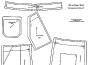
Once again, capricious and changeable fashion has taken its turn and reached the peak of popularity...

